GI Embryology: Foregut, Midgut & Hindgut
A Step 1 walkthrough of GI development — how the gut tube is divided into foregut, midgut, and hindgut by blood supply, what each segment becomes, and the four morphogenetic events (recanalization, herniation/rotation, pancreatic fusion, cloacal partition) whose failures produce the board-classic malformations: TEF, duodenal atresia, malrotation/volvulus, omphalocele vs gastroschisis, Meckel, and Hirschsprung — each with mechanism and presentation.
The gut tube and its three divisions
The primitive gut tube forms in week 4 as craniocaudal and lateral body folding incorporates the dorsal part of the endoderm-lined yolk sac into the embryo, producing a tube lined by endoderm (epithelium and glands) and wrapped in splanchnic mesoderm (smooth muscle, connective tissue, serosa). It is divided into three segments by blood supply, not by organ — a distinction the boards love:
- Foregut → celiac trunk (T12)
- Midgut → superior mesenteric artery / SMA (L1)
- Hindgut → inferior mesenteric artery / IMA (L3)
The junctions are clinically load-bearing. The foregut–midgut boundary sits in the 2nd part of the duodenum at the major duodenal papilla (ampulla of Vater / bile duct entry); the midgut–hindgut boundary lies at the junction of the proximal 2/3 and distal 1/3 of the transverse colon (the SMA/IMA watershed near the splenic flexure). This gives you a free localizer: obstruction above the ampulla → non-bilious vomiting; below it → bilious vomiting.
Highest-yield derivatives
- Foregut endoderm → esophagus, stomach, duodenum proximal to the ampulla, plus the outpouchings: respiratory (lung) bud, liver, gallbladder, and pancreas (ventral + dorsal buds). Supplied by the celiac trunk.
- Spleen is a trap: it arises from mesoderm of the dorsal mesentery (dorsal mesogastrium), NOT foregut endoderm — but it is supplied by the celiac (splenic) artery.
- Midgut → duodenum distal to the ampulla, jejunum, ileum, cecum, appendix, ascending colon, proximal 2/3 of transverse colon. Supplied by the SMA.
- Hindgut → distal 1/3 transverse colon, descending + sigmoid colon, rectum, and anal canal above the pectinate line. Supplied by the IMA.
- Pectinate (dentate) line = the hindgut endoderm/ectoderm boundary. Above: visceral innervation, superior rectal artery (IMA), internal iliac nodes, columnar epithelium. Below: somatic innervation (inferior rectal/pudendal → painful), inferior rectal artery, superficial inguinal nodes, squamous epithelium.
- Localizer: bilious vomiting = obstruction distal to the ampulla (midgut and beyond); non-bilious = at or above the pylorus.
Highest-yield congenital defects (instant recall)
- Duodenal atresia → "double bubble" + bilious vomiting + Down syndrome (failed recanalization).
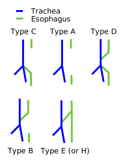
- Esophageal atresia + distal TEF (~85%) → polyhydramnios, coiled NG tube, gastric air bubble; screen VACTERL.
- Malrotation + midgut volvulus → bilious vomiting = surgical emergency (Ladd bands, incomplete 270° rotation).
- Hirschsprung → aganglionic rectum (always involved), no meconium in 48 h, Down / RET.
- Meckel diverticulum → painless GI bleed from ectopic gastric mucosa; persistent vitelline duct.
- Omphalocele (midline, covered, chromosomal) vs gastroschisis (right of umbilicus, uncovered, not chromosomal).
Segment → blood supply → derivatives
| Gut segment | Artery (vertebral level) | Endodermal derivatives |
|---|---|---|
| Foregut | Celiac trunk (T12) | Esophagus, stomach, duodenum (proximal to ampulla), liver, gallbladder, pancreas, respiratory bud |
| Midgut | SMA (L1) | Duodenum (distal to ampulla), jejunum, ileum, cecum, appendix, ascending colon, proximal 2/3 transverse colon |
| Hindgut | IMA (L3) | Distal 1/3 transverse colon, descending + sigmoid colon, rectum, upper anal canal (above pectinate line) |
Four morphogenetic events — and their signature failures
Most GI birth defects tested on Step 1 are failures of four dynamic developmental events. Learn the event and the malformation writes itself:
- Duodenal recanalization. The duodenal lumen is transiently obliterated by proliferating epithelium (~weeks 5–6) and must recanalize (~weeks 8–10). Failure → duodenal atresia/stenosis.
- Physiologic midgut herniation & rotation. Around week 6 the rapidly elongating midgut loop herniates through the umbilical ring; it rotates a total of 270° counterclockwise around the SMA axis (90° while herniated, then 180° on return) and retracts by ~week 10. Failure to return → omphalocele; incomplete/abnormal rotation → malrotation ± midgut volvulus with obstructing Ladd bands.
- Pancreatic bud fusion. The ventral bud swings posteriorly around the duodenum to fuse with the dorsal bud, and their ducts join. Abnormal ventral migration encircling the duodenum → annular pancreas; failure of the two ducts to fuse → pancreas divisum.
- Cloacal partition. The urorectal septum divides the cloaca into an anterior urogenital sinus and a posterior anorectal canal, and the anal membrane then perforates. Errors → imperforate anus / anorectal malformations.
Two more high-yield failures don't fit the four but are pure Step 1: failure of neural crest cell migration into the distal hindgut wall → Hirschsprung disease, and persistence of the vitelline (omphalomesenteric) duct → Meckel diverticulum.

Defect → mechanism → presentation
| Defect | Mechanism | Classic presentation |
|---|---|---|
| Esophageal atresia + distal TEF (~85%) | Faulty division of foregut by the tracheoesophageal septum | Polyhydramnios; drooling, choking/cyanosis with feeds; NG tube coils in blind pouch; gastric air bubble; screen VACTERL |
| Duodenal atresia | Failed recanalization of the duodenum | Bilious vomiting; "double bubble" x-ray; polyhydramnios; Down syndrome |
| Jejunal/ileal ("apple-peel") atresia | Vascular accident (SMA branch occlusion) in utero | Bilious vomiting; assoc. maternal smoking/cocaine (vasoconstriction) |
| Hypertrophic pyloric stenosis (postnatal, not embryologic) | Postnatal hypertrophy of pyloric muscularis | 2–6 wk firstborn male; projectile non-bilious vomiting; palpable "olive"; hypochloremic hypokalemic metabolic alkalosis; assoc. macrolides |
| Annular pancreas | Ventral bud encircles the 2nd duodenum | Duodenal obstruction; can mimic a double bubble |
| Pancreas divisum | Dorsal + ventral ducts fail to fuse | Usually asymptomatic; may cause pancreatitis |
| Omphalocele | Midgut fails to return to abdomen | Midline, herniation covered by peritoneum + amnion; assoc. trisomies 13/18/21, Beckwith-Wiedemann |
| Gastroschisis | Failure of lateral body-wall fold closure | Para-umbilical (usually right), no covering membrane; NOT chromosome-linked |
| Meckel diverticulum | Persistent vitelline duct (true diverticulum) | Painless lower-GI bleed/melena (ectopic gastric mucosa); pertechnetate (Meckel) scan; "Rule of 2s" |
| Malrotation ± volvulus | Incomplete 270° rotation | Bilious vomiting, Ladd bands, bowel ischemia |
| Hirschsprung disease | Failed neural crest migration → aganglionosis | No meconium in 48 h, distension; rectum always involved; RET; Down; suction biopsy |
| Imperforate anus | Abnormal urorectal septum / unperforated anal membrane | Absent anal opening ± fistula; VACTERL |
A term newborn develops coughing, choking, and cyanosis with the first feeding and has copious oral secretions (drooling). The pregnancy was complicated by polyhydramnios. A nasogastric tube cannot be advanced — it coils in the upper chest on film, yet the stomach is gas-filled.
Diagnosis: esophageal atresia with a distal tracheoesophageal fistula — the most common (~85%) type. The blind proximal pouch explains the drooling and coiled tube; the distal fistula lets air reach the stomach (and refluxed acid reach the lungs). The failed structure is the tracheoesophageal (respiratory) septum that should divide the foregut. Always screen for VACTERL. Contrast: pure atresia (no fistula) → gasless abdomen; H-type isolated fistula → recurrent aspiration pneumonias presenting later, not at the first feed.
A neonate with trisomy 21 features begins bilious vomiting within the first day of life; prenatal ultrasound had shown polyhydramnios. Abdominal x-ray shows the "double bubble" sign — a distended, air-filled stomach and proximal duodenum with no distal bowel gas.
Diagnosis: duodenal atresia from failed recanalization of the duodenum (~weeks 8–10). Because the block sits distal to the ampulla of Vater, the vomiting is bilious. High-yield association: Down syndrome. Note that an annular pancreas can produce an identical double bubble by extrinsically encircling the duodenum — same picture, different mechanism.
A full-term newborn fails to pass meconium within the first 48 hours, then develops abdominal distension and bilious vomiting. On digital rectal exam there is an explosive gush of gas and stool. Contrast enema shows a narrow distal segment with dilated proximal bowel (a transition zone).
Diagnosis: Hirschsprung disease — arrest of neural crest cell migration leaving the distal bowel aganglionic (absent Meissner submucosal and Auerbach myenteric plexuses), so it cannot relax. The rectum is ALWAYS involved, with disease extending a variable distance proximally. Associations: Down syndrome and RET proto-oncogene mutations. Gold-standard diagnosis: rectal suction biopsy showing absent ganglion cells; treatment resects the aganglionic segment.
- VACTERL — screen for these whenever you see TEF or imperforate anus: Vertebral defects, Anal atresia, Cardiac, Tracheo-Esophageal fistula, Renal, Limb.
- Meckel diverticulum — "Rule of 2s": ~2% of people, 2 feet from the ileocecal valve, 2 inches long, presents by age 2, 2:1 male, 2 ectopic tissue types (gastric + pancreatic), and ~2% become symptomatic.
- Midgut rotation = 270° counterclockwise around the SMA (90° while herniated + 180° on return) — hold "counterclockwise, as viewed from the front."
Practice Embryology now
Board-style questions, spaced-repetition flashcards, and a Socratic AI tutor — free to start.