Genital Development & Sexual Differentiation
A high-yield Step 1 walkthrough of sexual differentiation: the indifferent stage, the SRY switch, and the three hormones (AMH, testosterone, DHT) that build the male tract — with verified derivative and congenital-defect tables and paired CAIS vs. 5α-reductase vignettes.
The developmental sequence: one blueprint, two outcomes
Both sexes begin identical at the indifferent (bipotential) stage. The story is a sequence of switches, and for boards the default program is female — the male pathway must be actively imposed by the Y chromosome.
- Gonadal ridge forms from intermediate mesoderm (urogenital ridge) plus overlying coelomic epithelium. Primordial germ cells arise in the wall of the yolk sac and migrate along the dorsal mesentery of the hindgut to colonize the gonadal ridge (~weeks 4–6).
- Two duct systems develop in every embryo: the mesonephric (Wolffian) ducts and the paramesonephric (Müllerian) ducts.
- The switch: the SRY gene on the short arm of the Y chromosome encodes testis-determining factor (TDF), which drives the gonadal medulla to become a testis. No SRY → the cortex develops into an ovary.
- Hormones decide the ducts and the external genitalia (see next block). By the end of ~week 12 the external genitalia are distinguishable.
Everything the boards test is a variation on this theme: which switch failed, which duct persisted or regressed, and which hormone was missing.
- Female is the boards default. Female development needs neither ovary nor hormone — it is the absence of SRY, AMH, and testosterone.
- SRY (Y short arm) → TDF → testis (from the medulla). No SRY → ovary (from the cortex).
- Three testicular products: Sertoli cells → AMH (regress Müllerian ducts); Leydig cells → testosterone (maintain Wolffian ducts); testosterone → DHT via 5α-reductase (builds external genitalia + prostate).
- Testosterone → INTERNAL male ducts (SEED); DHT → EXTERNAL genitalia + prostate. This split is the key to 5α-reductase deficiency.
- Müllerian → fallopian tubes, uterus, cervix, upper vagina. Wolffian → SEED. Prostate and the lower vagina come from the UROGENITAL SINUS, not the ducts.
- Remnants tested: appendix of testis = Müllerian remnant (male); Gartner duct cyst = Wolffian (mesonephric) remnant (female).
- Highest-yield differentiation defects: CAIS (46,XY, dead androgen receptor → female phenotype, no uterus, testes); 5α-reductase deficiency (46,XY, no DHT → ambiguous at birth, virilize at puberty); MRKH/Müllerian agenesis (46,XX, no uterus/upper vagina, normal ovaries); CAH / 21-hydroxylase deficiency (46,XX, virilized female).
- Gonadal dysgenesis: streak gonads in Turner (45,X); seminiferous tubule dysgenesis in Klinefelter (47,XXY).
Indifferent structure → male vs. female derivative
| Indifferent / precursor | Male derivative | Female derivative |
|---|---|---|
| Gonad (medulla vs. cortex) | Testis (medulla) | Ovary (cortex) |
| Mesonephric (Wolffian) duct | Epididymis, ductus (vas) deferens, seminal vesicle, ejaculatory duct | Regresses — remnant = Gartner duct |
| Paramesonephric (Müllerian) duct | Regresses — remnant = appendix of testis | Fallopian tubes, uterus, cervix, upper vagina |
| Urogenital sinus | Bladder, prostate, prostatic urethra, bulbourethral (Cowper) glands | Bladder, urethra, lower vagina, greater vestibular (Bartholin) & paraurethral (Skene) glands |
| Genital tubercle | Glans penis, corpora cavernosa/spongiosum | Glans clitoris |
| Urogenital (urethral) folds | Ventral shaft + penile (spongy) urethra | Labia minora |
| Labioscrotal swellings | Scrotum | Labia majora |
| Gubernaculum | Gubernaculum testis (anchors descended testis) | Ovarian ligament + round ligament of uterus |
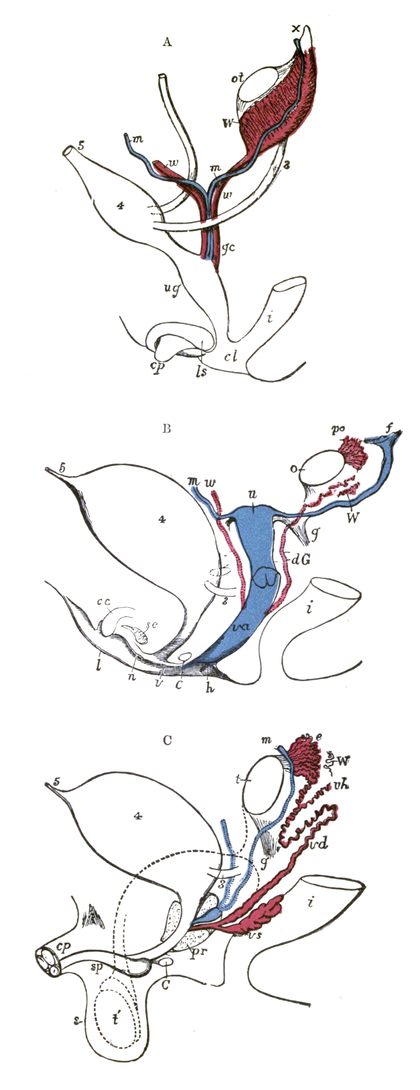
Who runs the program: three hormones
Male differentiation is built by three signals, each with a distinct job — and each with its own classic disease when it fails:
- Sertoli cells → Anti-Müllerian Hormone (AMH / MIF). AMH causes regression of the Müllerian ducts locally. Without it, a genetic male retains a uterus and tubes (persistent Müllerian duct syndrome).
- Leydig cells → testosterone. Testosterone acts as a paracrine signal that maintains and virilizes the Wolffian ducts into the internal male structures (SEED).
- DHT (dihydrotestosterone). Target tissues convert testosterone to DHT via 5α-reductase; DHT builds the external genitalia and the prostate (penis, scrotum, urethral fusion).
The female default: no SRY → no testis → no AMH (Müllerian ducts persist) and no testosterone (Wolffian ducts regress). A functioning ovary is not required to form the female internal tract — which is exactly why Müllerian agenesis and androgen insensitivity look the way they do.
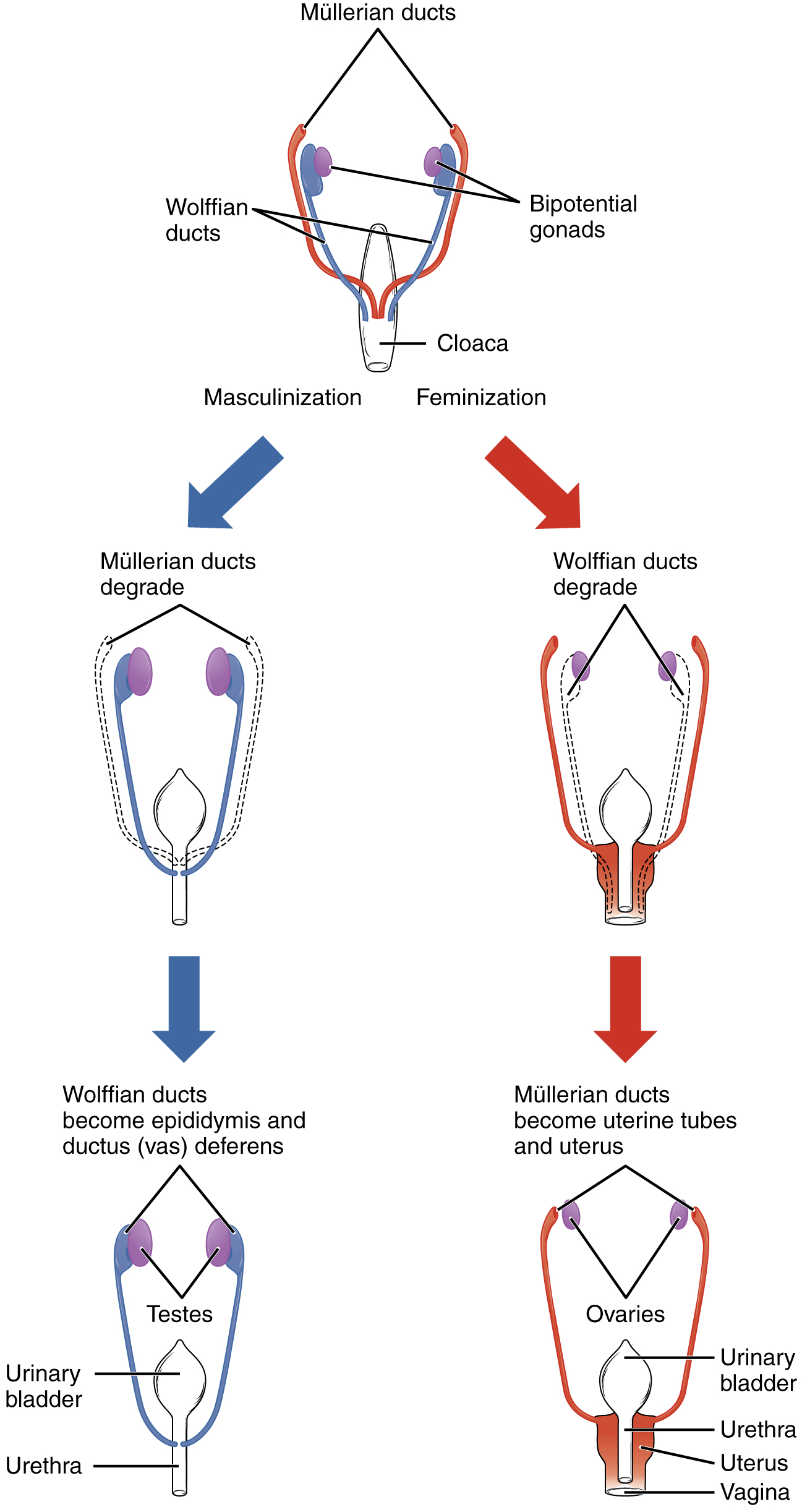
Wolffian (mesonephric) duct → SEED:
- S — Seminal vesicles
- E — Epididymis
- E — Ejaculatory duct
- D — Ductus (vas) deferens
Trap: the prostate is NOT in SEED — it comes from the urogenital sinus (DHT-driven).
Müllerian (paramesonephric) duct → the female tract: Fallopian tubes, Uterus, Cervix, Upper vagina.
Pairing hormone → duct:
- Sertoli → AMH → Abolishes Müllerian ducts (AMH = Anti-Müllerian Hormone).
- Leydig → Testosterone → maintains Wolffian ducts (the SEED).
A 16-year-old presents for primary amenorrhea. She has normal breast development (Tanner IV) but scant axillary and pubic hair. Exam reveals a short, blind-ending vagina with no palpable cervix or uterus; there are bilateral inguinal masses. Imaging confirms an absent uterus and ovaries. Karyotype is 46,XY; serum testosterone is in the normal male range, with elevated LH and estrogen.
Diagnosis: Complete Androgen Insensitivity Syndrome (CAIS) — a loss-of-function mutation in the androgen receptor (X-linked recessive).
Why it looks like this:
- Testes make AMH → Müllerian ducts regress → no uterus/tubes/upper vagina (blind vaginal pouch).
- Testes make testosterone, but receptors are blind to it → Wolffian ducts don't develop and the external genitalia stay female.
- Testosterone is aromatized to estrogen → breast development at puberty; androgen-dependent hair is scant.
Pearl: the inguinal masses are testes — gonadectomy is planned because of malignancy risk (typically deferred until after puberty to allow spontaneous feminization).
Classic congenital defects → mechanism → presentation
| Defect | Mechanism | Karyotype | Presentation |
|---|---|---|---|
| Complete androgen insensitivity (CAIS) | Nonfunctional androgen receptor; AMH still regresses Müllerian ducts, testosterone can't act → female external genitalia; T aromatized to estrogen | 46,XY | Phenotypic female, breasts, primary amenorrhea, blind vagina, no uterus, scant hair, testes in inguinal canal; ↑T, ↑LH |
| 5α-reductase deficiency | Cannot convert T → DHT; DHT-dependent external genitalia under-virilized until the pubertal testosterone surge | 46,XY (AR) | Ambiguous/female-appearing at birth, virilizes at puberty ("penis at 12"); normal internal male ducts; ↑ T:DHT ratio |
| Müllerian agenesis (MRKH) | Failure of paramesonephric duct development | 46,XX | Primary amenorrhea, absent uterus & upper vagina, normal ovaries & secondary sex characteristics; often renal anomalies |
| Müllerian fusion/resorption defects (bicornuate, uterus didelphys, septate uterus) | Failure of the paired Müllerian ducts to fuse (didelphys, bicornuate) or of the median septum to resorb (septate) | 46,XX | Often silent; recurrent pregnancy loss, preterm labor; associated renal/urinary anomalies |
| Hypospadias | Failure of the urogenital (urethral) folds to fuse (inadequate DHT); more common than epispadias | — | Urethral opening on the ventral penis |
| Epispadias | Faulty positioning of the genital tubercle | — | Urethral opening on the dorsal penis; associated with bladder exstrophy |
| Congenital adrenal hyperplasia (21-hydroxylase def.) | Excess adrenal androgens virilize a female fetus | 46,XX | Ambiguous/virilized external genitalia, clitoromegaly; normal uterus/ovaries; salt-wasting in classic form |
| Cryptorchidism | Failure of testicular descent (gubernaculum / hormonal) | 46,XY | Undescended testis; ↑ risk of germ cell tumor and infertility |
A 13-year-old raised as a girl is brought in for progressive virilization at puberty: deepening voice, phallic (clitoral) enlargement, increased muscle mass, and testicular descent into the labioscrotal folds. She was born with ambiguous genitalia. Karyotype is 46,XY; testosterone is normal, DHT is low, and the testosterone:DHT ratio is elevated. No uterus is present on imaging.
Diagnosis: 5α-reductase deficiency (autosomal recessive).
Contrast with CAIS — this is the high-yield pair:
- Internal ducts are normal male (Wolffian derivatives) because they depend on testosterone, which is present and active (the receptor works here).
- External genitalia depend on DHT → under-virilized at birth → ambiguous.
- No uterus, because Sertoli AMH still regressed the Müllerian ducts.
- At puberty, the surge in testosterone partially compensates → virilization occurs.
Key discriminator: in 5α-reductase deficiency the patient virilizes at puberty; in CAIS the androgen receptor is broken, so virilization never happens and estrogen effects (breasts) dominate.
Practice Embryology now
Board-style questions, spaced-repetition flashcards, and a Socratic AI tutor — free to start.