Fever of Unknown Origin
A boards-focused Step 2 CK lesson on fever of unknown origin: the Petersdorf/Durack definitions and four etiologic buckets, exposure-driven organism matching, and next-best-step decisions anchored by classic vignettes (giant cell arteritis, adult-onset Still disease) and the staged diagnostic ladder ending in FDG-PET/CT and directed biopsy.
What Counts as FUO — and the Big Four Buckets
Fever of unknown origin (FUO) is classically defined (Petersdorf & Beeson) as T >38.3°C (101°F) on several occasions, lasting >3 weeks, undiagnosed after an appropriate initial evaluation (originally 1 week in-hospital; modern practice accepts ≥3 outpatient visits or 3 inpatient days). Durack & Street split FUO into four types — classic, nosocomial, neutropenic (immunodeficient), and HIV-associated — each with its own differential.
Four etiologic buckets drive the classic workup:
- Infection (~20–40%): TB (miliary/extrapulmonary), occult abscess, culture-negative endocarditis, osteomyelitis, EBV/CMV, HIV.
- Malignancy (~10–30%): lymphoma (esp. non-Hodgkin), leukemia, renal cell carcinoma, atrial myxoma.
- Non-infectious inflammatory (~15–30%): adult-onset Still disease, SLE, giant cell arteritis, polyarteritis nodosa, IBD.
- Miscellaneous/undiagnosed: drug fever, factitious fever, familial Mediterranean fever, subacute thyroiditis, VTE.
Age skews the odds: elderly → giant cell arteritis and malignancy; younger adults → infection and inflammatory disease. Up to ~30% remain undiagnosed — and these often resolve spontaneously with a good prognosis.
- Draw ≥3 blood cultures OFF antibiotics before any empiric therapy — premature antibiotics convert true endocarditis into "culture-negative."
- Stop every nonessential drug. Drug fever is classically a well-appearing patient with relative bradycardia and eosinophilia that resolves within ~72h after withdrawal. Culprits: beta-lactams, sulfonamides, anticonvulsants, allopurinol.
- Do NOT give empiric antibiotics or steroids to a stable patient — they mask the diagnosis. Exception: suspected GCA → start steroids immediately to prevent blindness.
- FDG-PET/CT is the high-yield next step when history, exam, first-tier labs, and CT are unrevealing — it localizes occult infection, large-vessel vasculitis, and malignancy to direct biopsy.
- Repeat the history and exam frequently; a new murmur, node, rash, or tender temporal artery emerges over time and cracks the case.
- Very high ferritin + salmon rash + arthralgia + sore throat = adult-onset Still disease.
- Pulse–temperature dissociation (Faget sign) narrows to typhoid, Legionella, Q fever, RMSF/typhus, leptospirosis, and drug/factitious fever.
Vignette: A 74-year-old woman has 4 weeks of fever, fatigue, and 5 kg of weight loss. She now reports a new bitemporal headache, scalp tenderness when combing her hair, and jaw pain while chewing. Exam reveals a tender, nodular, pulseless temporal artery. ESR is 92 mm/hr and CRP is markedly elevated.
- Diagnosis: Giant cell (temporal) arteritis — a leading cause of FUO in patients >50.
- Next best step: Start high-dose corticosteroids IMMEDIATELY — do not wait for biopsy. Vision loss from arteritic anterior ischemic optic neuropathy is sudden and irreversible but preventable.
- Confirm with: temporal artery biopsy within ~1–2 weeks (remains positive despite early steroids); temporal/axillary ultrasound (halo sign) is an adjunct.
- Associated: polymyalgia rheumatica (shoulder/hip-girdle stiffness) in ~40–50%.
- Board trap: ordering the biopsy before steroids in a patient with visual symptoms — the biopsy must never delay treatment.
Vignette: A 29-year-old man has 5 weeks of daily fevers spiking to 39.5°C that return to baseline between spikes (quotidian). With each spike he develops a transient salmon-colored maculopapular rash on the trunk, arthralgias, and a sore throat. Labs: WBC 18,000 (neutrophil-predominant), ferritin 9,000 ng/mL, ANA and RF negative, blood cultures sterile.
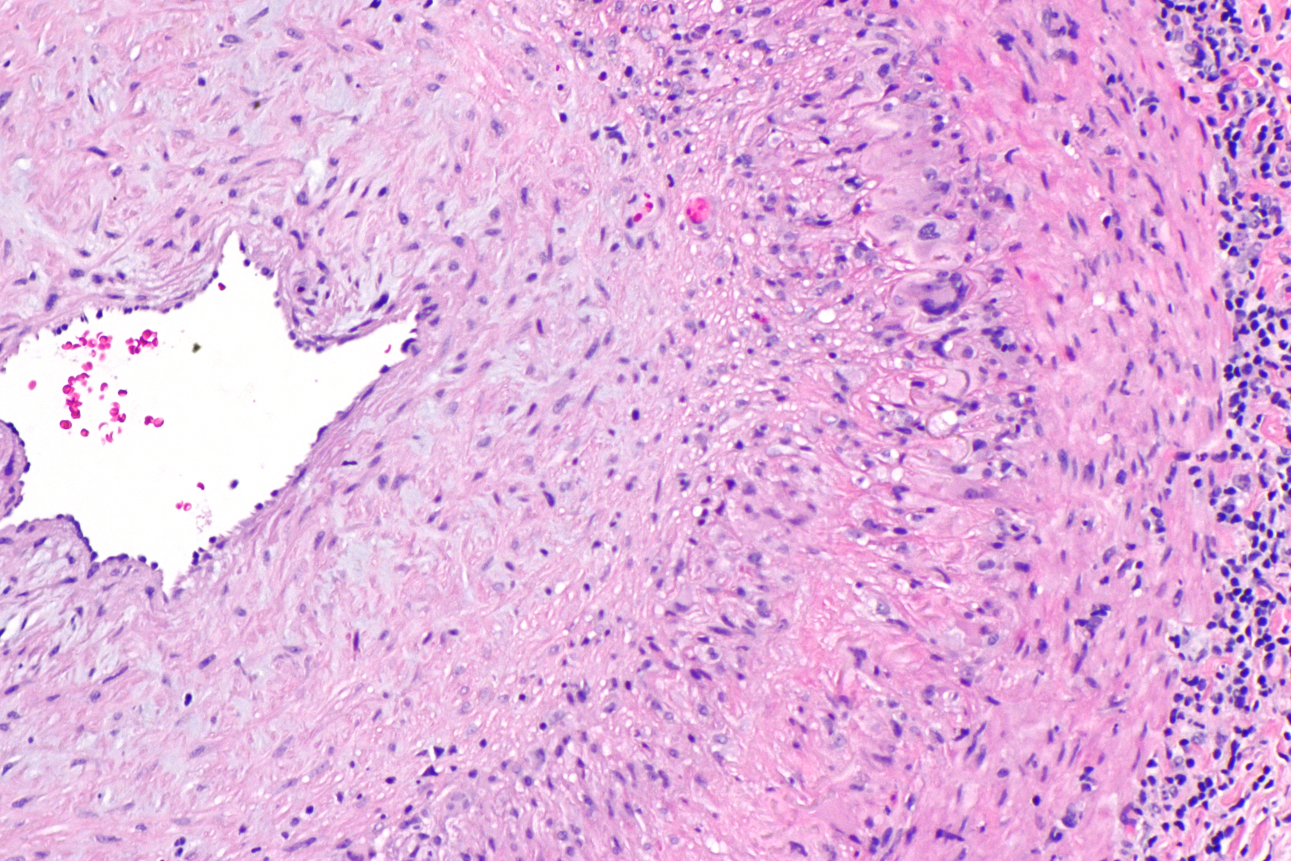
- Diagnosis: Adult-onset Still disease (Yamaguchi criteria) — a diagnosis of exclusion.
- Clues: quotidian fever, evanescent rash appearing with the fever spike, extreme hyperferritinemia (with a low glycosylated fraction), leukocytosis, seronegative serologies.
- Next best step: exclude infection and malignancy first (cultures, imaging ± marrow), then treat with NSAIDs, escalating to corticosteroids ± IL-1/IL-6 blockade (anakinra, tocilizumab).
- Watch for: macrophage activation syndrome (worsening cytopenias, falling ESR with rising ferritin) — a life-threatening complication.
Match the Exposure — Zoonotic & Travel FUO
| Exposure clue | Organism (disease) | Buzzword | First-line treatment |
|---|---|---|---|
| Parturient livestock/cats; culture-neg endocarditis | Coxiella burnetii (Q fever) | Pulse–temp dissociation, granulomatous hepatitis | Doxycycline (+ hydroxychloroquine if chronic) |
| Unpasteurized dairy, goats/cattle | Brucella | Undulant fever, sacroiliitis | Doxycycline + rifampin (or doxycycline + aminoglycoside) |
| Cat scratch/bite | Bartonella henselae | Tender regional lymphadenopathy | Azithromycin; doxy+rifampin if systemic |
| Travel to S. Asia, fecal–oral | Salmonella Typhi | Rose spots, Faget sign, stepwise fever | Ceftriaxone or azithromycin |
| Freshwater / rodent urine | Leptospira | Conjunctival suffusion, Weil (jaundice+AKI) | Doxy (mild); IV penicillin/ceftriaxone (severe) |
| Tick bite, SE/S-central US | Rickettsia rickettsii (RMSF) | Rash wrists/ankles → centripetal, palms/soles | Doxycycline (all ages) |

Faget sign = relative bradycardia: the heart rate is inappropriately low for the height of the fever. Judge it only when the temperature is genuinely high — apply the sign only above 38.9°C (102°F), and only in a patient not on a beta-blocker/calcium-channel blocker and without heart block or a pacemaker (an intact conduction system is required). As a rule of thumb (Cunha), each 1°F of fever adds roughly 10 bpm, so at 39–40°C the pulse should be ~110–130 — a pulse under 100 is the classic tell.
There is no tidy acronym; the highest-yield move is to group the causes by category:
- Intracellular / atypical bacteria: Legionella, Coxiella burnetii (Q fever), Salmonella Typhi (typhoid), Chlamydia psittaci (psittacosis)
- Rickettsial: Rocky Mountain spotted fever and typhus
- Spirochete: leptospirosis (the classic extracellular exception)
- Protozoal: malaria and babesiosis
- Non-infectious mimics: drug fever and factitious fever (the reading is manipulated, so there is no real tachycardia), plus CNS lesions / raised intracranial pressure
Pattern: mostly intracellular/atypical organisms — plus the drug/factitious-fever mimics you must not miss.
The Staged Workup — Know the Order of Next Steps
Boards test the sequence. Work up cost-consciously from the least to the most invasive:
- History & exam, repeated. Chase travel, animal/occupational exposures, sick contacts, TB risk, sexual history, all drugs/supplements, family history (FMF), and prior surgery/prosthetic hardware.
- Discontinue nonessential medications (drug fever).
- First-tier labs: CBC with differential + peripheral smear, CMP/LFTs, ESR & CRP, LDH, ferritin, ≥3 blood cultures off antibiotics, urinalysis + culture, HIV, TB testing (IGRA), ANA/RF, and a chest X-ray.
- Cross-sectional imaging: CT chest/abdomen/pelvis for abscess, adenopathy, or tumor; echocardiography (TTE → TEE) if endocarditis is suspected.
- FDG-PET/CT when the above is unrevealing — the best functional test to localize an inflammatory, malignant, or infectious focus.
- Directed biopsy of the abnormal site (lymph node, liver, temporal artery, bone marrow).
- Empiric therapy only if the patient is deteriorating — otherwise observe; many undiagnosed FUOs are benign and self-limited.
- HIV-associated FUO: disseminated MAC, TB, histoplasmosis, CMV, lymphoma, or IRIS — check the CD4 count, which reframes the entire differential.
- Neutropenic FUO: assume infection (gram-negative bacteremia, invasive fungal — Candida, Aspergillus). Start empiric broad-spectrum antibiotics immediately — this is the one setting where you do NOT wait.
- Nosocomial FUO: think C. difficile, catheter/line infection, drug fever, DVT/PE, NG-tube sinusitis, and acalculous cholecystitis.
- Factitious fever: young, healthcare-associated patient; absent diurnal variation and pulse–temperature discordance; very high "temps" without tachycardia or diaphoresis — witness the measurement and check a simultaneous fresh-void urine temperature.
- Familial Mediterranean fever: recurrent short febrile serositis + Mediterranean ancestry → colchicine.
- VTE (DVT/PE): an under-recognized non-infectious cause of persistent fever.
Practice Infectious Disease now
Board-style questions, spaced-repetition flashcards, and a Socratic AI tutor — free to start.