Face, Palate & Cleft Development
A high-yield Step 1 walkthrough of face development (weeks 4–8, palate completing by ~week 12) from the five neural-crest-derived facial prominences and their derivatives, then the boards-favorite congenital clefts and first-arch anomalies — cleft lip vs. cleft palate, median/oblique clefts, and Pierre Robin sequence — each tied to its fusion-failure mechanism.
Building the Face: Weeks 4–8
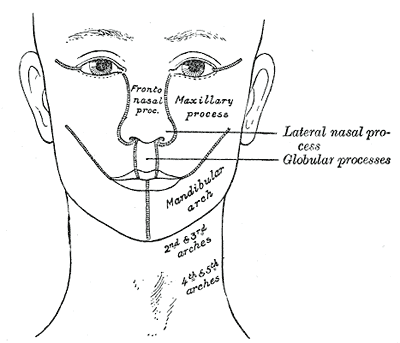
The face forms between weeks 4 and 8 from five mesenchymal swellings ("prominences") that surround the primitive mouth (stomodeum). Critically, the mesenchyme of every facial prominence is neural crest–derived (ectomesenchyme). The five prominences are:
- One unpaired frontonasal prominence (superior/midline)
- Two maxillary prominences
- Two mandibular prominences
The maxillary and mandibular prominences both arise from the first pharyngeal arch. Ectodermal thickenings called nasal placodes appear on the frontonasal prominence, invaginate into nasal pits, and become flanked by horseshoe-shaped medial and lateral nasal prominences. The upper lip is completed when the two maxillary prominences grow medially and push the two medial nasal prominences toward the midline, where the medial nasal prominences merge to form the intermaxillary segment. The facial clefts tested on the boards are almost always a failure of fusion/merging of these prominences — not tissue splitting apart.
- Five prominences: 1 frontonasal + 2 maxillary + 2 mandibular (maxillary & mandibular = 1st pharyngeal arch); all mesenchyme is neural crest.
- Medial nasal prominences merge → intermaxillary segment → philtrum of upper lip, primary palate (premaxilla), the 4 maxillary incisors, and the crest/tip of the nose.
- Lateral nasal prominences → alae (sides/wings) of the nose — not the lip.
- Maxillary prominences → lateral upper lip + cheeks, most of the maxilla, and the secondary palate (lateral palatine shelves).
- Mandibular prominences → lower lip, mandible, chin.
- Upper lip = medial nasal (philtrum) + maxillary (lateral parts); lower lip = mandibular.
- Nasolacrimal duct develops in the groove between the lateral nasal and maxillary prominences.
- Incisive foramen = the dividing landmark: primary palate/lip anterior, secondary palate posterior.

Structure → Derivative
| Embryonic structure | Adult derivative(s) |
|---|---|
| Frontonasal prominence | Forehead, bridge/dorsum & root of nose; source of the medial & lateral nasal prominences |
| Medial nasal prominences (→ intermaxillary segment) | Philtrum of upper lip; crest & tip of nose; primary palate (premaxilla) + 4 incisors |
| Lateral nasal prominences | Alae (sides) of the nose |
| Maxillary prominences | Lateral upper lip, cheeks, maxilla, secondary palate |
| Mandibular prominences | Lower lip, mandible, chin |
| Nasolacrimal groove (lateral nasal–maxillary) | Nasolacrimal duct & lacrimal sac |
Making the Palate: Primary vs. Secondary
The palate has two embryologic parts divided by the incisive foramen.
- Primary palate (median palatine process): derives from the intermaxillary segment and forms the small premaxillary wedge anterior to the incisive foramen that carries the four incisors.
- Secondary palate: forms the hard palate behind the foramen plus the soft palate, and derives from the two lateral palatine shelves of the maxillary prominences.
Sequence of secondary palate closure:
- The palatine shelves first grow vertically, downward alongside the developing tongue.
- Around week 7, as the mandible enlarges and the tongue descends, the shelves elevate to the horizontal and grow toward the midline.
- They then fuse with each other, with the primary palate anteriorly, and with the down-growing nasal septum superiorly, completing by roughly weeks 10–12.
Key mechanistic point: anything that keeps the tongue high (e.g., micrognathia) mechanically blocks shelf elevation → cleft palate.
Defect → Mechanism
| Malformation | Failed fusion / mechanism | Key features |
|---|---|---|
| Cleft lip (± primary palate) | Maxillary fails to fuse with medial nasal prominence (intermaxillary segment) | Cleft in upper lip lateral to midline (philtral column), anterior to incisive foramen; more common in males; uni- or bilateral |
| Cleft palate | Palatine shelves fail to fuse with each other / primary palate / nasal septum | Defect posterior to incisive foramen; more common in females; bifid uvula = mildest form |
| Median cleft lip | Two medial nasal prominences fail to merge | Midline upper-lip cleft; assoc. holoprosencephaly (hypotelorism) and trisomy 13 (Patau) |
| Oblique facial cleft | Maxillary fails to fuse with lateral nasal prominence | Cleft running from lip to eye along an exposed nasolacrimal duct |
| Congenital nasolacrimal duct obstruction | Nasolacrimal cord fails to canalize | Neonatal epiphora (tearing), recurrent dacryocystitis |
A newborn has a strikingly small, receding lower jaw and difficulty feeding with intermittent airway obstruction that worsens supine and improves prone. Oral exam shows a wide U-shaped cleft of the soft and hard palate, and the tongue falls posteriorly.
This is Pierre Robin sequence. A primary defect of mandibular hypoplasia (micrognathia) forces the tongue to sit high and posteriorly (glossoptosis), which mechanically prevents the palatine shelves from elevating and fusing → cleft palate. It is a true "sequence": one initiating anomaly cascades into the others, and this is why airway/feeding management (prone positioning) is central.
Contrast — Treacher Collins syndrome (autosomal dominant, TCOF1): a neural-crest deficiency of first-arch derivatives producing mandibular + zygomatic (malar) hypoplasia, downslanting palpebral fissures, lower-eyelid colobomas, and external ear anomalies (often conductive hearing loss).
- Incisive foramen = the divider. Everything a cleft Lip involves (lip, premaxilla, incisors) is anterior; the secondary Palate is posterior. Anterior = Lip, Posterior = Palate.
- Pierre Robin cascade — run it in order: Micrognathia → Glossoptosis → Cleft palate (small jaw pushes the tongue back and up; the tongue blocks the shelves).
- L for Lateral = aLae (wings/sides of nose); M for Medial = Middle (philtrum/premaxilla). Keeps the two nasal prominences straight.
- Two distinct groups: cleft lip ± palate (more common overall, more in males) vs. isolated cleft palate (more common in females).
- Bifid uvula = mildest form of (secondary) cleft palate.
- Multifactorial etiology: risk ↑ with maternal smoking, anticonvulsants (valproate; phenytoin → fetal hydantoin syndrome), alcohol, corticosteroids, and folate deficiency; periconceptional folate lowers risk.
- Complications of cleft palate: feeding difficulty, nasal regurgitation, recurrent otitis media (tensor veli palatini/Eustachian-tube dysfunction), and speech problems (velopharyngeal insufficiency).
- Syndromic associations to know: Pierre Robin sequence, Treacher Collins, DiGeorge/22q11.2 (cleft palate + velopharyngeal insufficiency), trisomy 13 (median cleft lip, holoprosencephaly).
Practice Embryology now
Board-style questions, spaced-repetition flashcards, and a Socratic AI tutor — free to start.