Epithelia & Cell Junctions
A board-focused tour of epithelial classification and the epithelial junctional complex — tight, adherens, desmosome, and gap junctions plus hemidesmosomes — mapping each junction's protein components to the high-yield blistering and genetic diseases (pemphigus vs. bullous pemphigoid, connexin mutations, E-cadherin loss) tested on Step 1.
Epithelia: the polarized barrier cells
Epithelia are avascular sheets of tightly packed, polarized cells that cover surfaces, line cavities, and form glands. They sit on a basement membrane, are nourished by diffusion from the underlying connective tissue, and regenerate rapidly. Every epithelial cell has three domains — an apical surface (facing the lumen or outside world), lateral surfaces (cell-to-cell contact), and a basal surface (facing the basement membrane). Cell junctions knit these cells together, seal the paracellular space, anchor the sheet to matrix, and let cells talk to one another. Epithelia are classified two ways: by the number of cell layers (simple = one, stratified = many) and by cell shape (squamous, cuboidal, columnar).
- Simple squamous — alveoli, endothelium (vessels), mesothelium (serous cavities), Bowman capsule; built for diffusion and filtration
- Simple cuboidal — kidney tubules, thyroid follicles, small ducts
- Simple columnar — GI tract (stomach → rectum), gallbladder, uterus
- Pseudostratified ciliated columnar — respiratory tract, fallopian tube; every cell touches the basement membrane but nuclei sit at different heights, so it only looks layered
- Stratified squamous, keratinized — epidermis; non-keratinized — oral mucosa, esophagus, vagina, anal canal (distal)
- Transitional (urothelium) — bladder, ureter, renal pelvis; stretches with distension
- Epithelium is cytokeratin-positive (a carcinoma marker); mesenchymal cells are vimentin-positive

The junctional complex, apical to basal
Moving from the apical surface downward, epithelial cells share a stereotyped arrangement of junctions. Strictly, the classic apical junctional complex is the top three (tight → adherens → desmosome); gap junctions sit lower, scattered along the lateral membrane:
- Tight junction (zonula occludens) — the most apical junction; claudins and occludins fuse adjacent membranes to block paracellular leak (the gate) and to fence apical membrane proteins away from basolateral ones, maintaining cell polarity (the fence).
- Adherens junction (zonula adherens) — a continuous belt of E-cadherin (Ca²⁺-dependent) tied to the actin cytoskeleton through catenins.
- Desmosome (macula adherens) — 'spot welds' of desmoglein and desmocollin anchored to keratin intermediate filaments via plakoglobin and desmoplakin.
- Gap junction — connexons (each built from six connexins) form pores allowing direct passage of ions and small molecules (<~1 kDa), enabling electrical and metabolic coupling.
Anchoring the entire sheet to the basement membrane are hemidesmosomes, where integrins bind laminin and connect back to keratin filaments (a cell-to-matrix junction, not cell-to-cell).
Junctions at a glance
| Junction (apical → basal) | Key proteins | Cytoskeletal link | Function |
|---|---|---|---|
| Tight junction (zonula occludens) | Claudins, occludins | Actin (ZO scaffold) | Paracellular barrier + polarity |
| Adherens junction (zonula adherens) | E-cadherin, catenins | Actin | Cell–cell adhesion belt |
| Desmosome (macula adherens) | Desmoglein, desmocollin | Keratin (intermediate filaments) | Spot-weld cell–cell adhesion |
| Gap junction | Connexons / connexins | — | Communication (ions, molecules <~1 kDa) |
| Hemidesmosome | Integrins (bind laminin) | Keratin | Anchors cell to basement membrane |
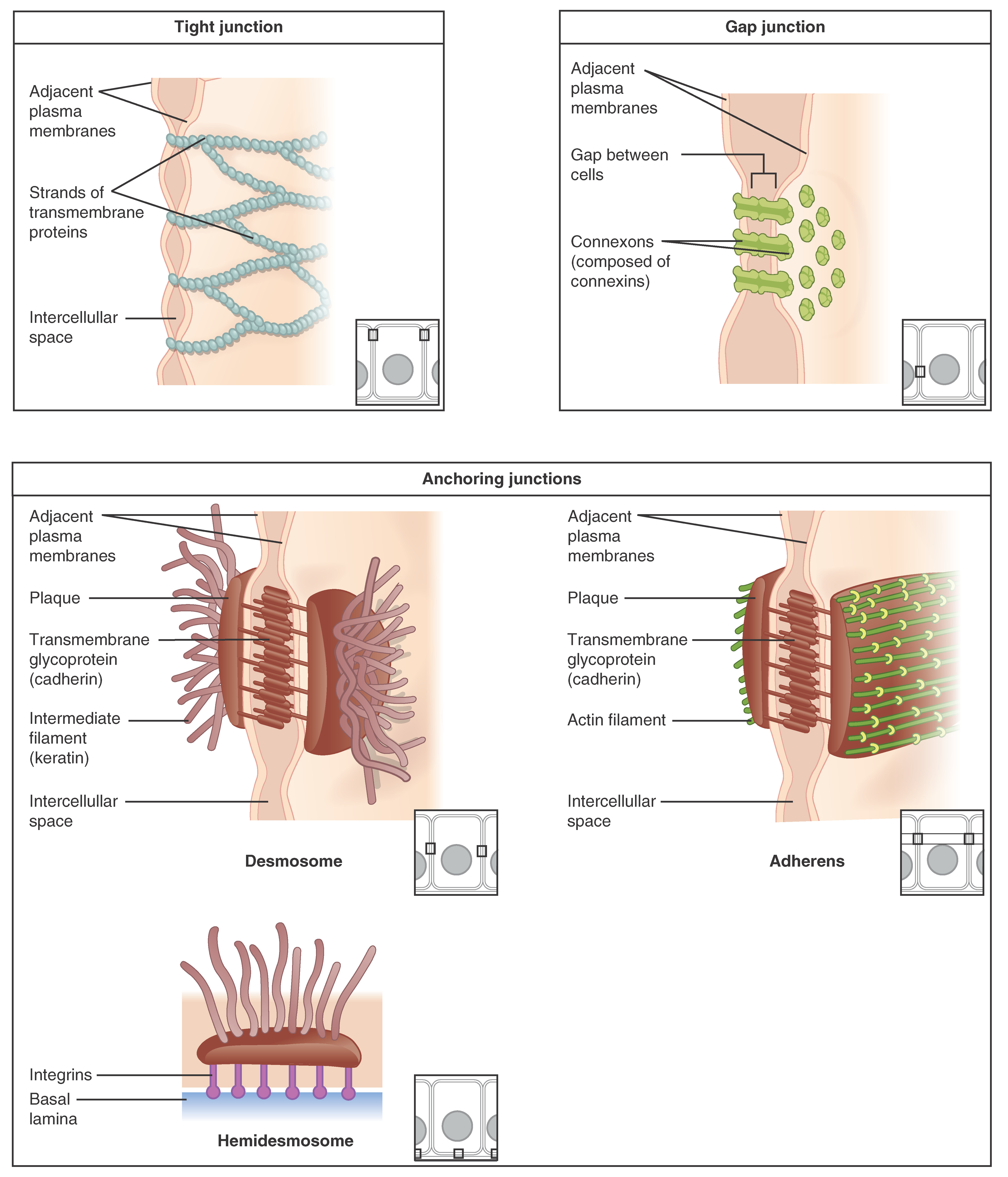
- Tight junction = claudins + occludins → paracellular barrier and apical–basal polarity; also form the blood–brain barrier (capillary endothelium) and blood–testis barrier (Sertoli cells)
- Adherens junction and desmosome use cadherins (Ca²⁺-dependent); adherens links to actin, desmosome links to keratin
- Gap junction = connexons built from connexins; couple cells electrically (e.g., cardiac myocytes propagate action potentials via connexin-43 in intercalated discs)
- Hemidesmosome = integrins → laminin in the basal lamina (cell-to-matrix)
- Basement membrane = basal lamina (type IV collagen + laminin) + reticular lamina; type IV collagen is the antigen in Goodpasture and the mutated protein in Alport
- Connexin 26 (GJB2) → congenital sensorineural deafness; connexin 32 (GJB1) → X-linked Charcot-Marie-Tooth
A blistering skin disease is a junction disease — match the antibody to the junction.
- Pemphigus vulgaris — IgG against desmoglein-3 (± desmoglein-1) in desmosomes → acantholysis and intraepidermal (suprabasal), flaccid bullae; Nikolsky sign positive; painful oral erosions (often the first sign); biopsy shows a 'row of tombstones,' and immunofluorescence shows a reticular / 'fishnet' intercellular pattern.
- Bullous pemphigoid — IgG against hemidesmosomes (BP180 / BP230) → subepidermal, tense bullae in the elderly; Nikolsky negative; oral mucosa usually spared; linear IgG/C3 along the basement membrane.
Loss of E-cadherin promotes invasion and metastasis: CDH1 mutation underlies diffuse (signet-ring) gastric carcinoma and lobular breast carcinoma. On boards, a named junction-protein defect points straight to its disease.
Pemphigus vulgaris vs. bullous pemphigoid
| Feature | Pemphigus vulgaris | Bullous pemphigoid |
|---|---|---|
| Target | Desmoglein (desmosome) | Hemidesmosome (BP180/BP230) |
| Blister plane | Intraepidermal (suprabasal) | Subepidermal |
| Bullae | Flaccid | Tense |
| Nikolsky sign | Positive | Negative |
| Oral involvement | Common (often first) | Usually spared |
| Immunofluorescence | Reticular / fishnet (intercellular) | Linear (basement membrane) |
| Course | More severe | Milder, older patients |
Bullous Pemphigoid = the 'B' disease:
- Below the epidermis (subepidermal split)
- Basement membrane / hemidesmosome target
- Bigger, tense Bullae; Nikolsky negative; Benign-er (milder) course
By contrast, Pemphigus vulgaris is more superficial (intraepidermal), flaccid, and Nikolsky Positive — 'PV = Positive.'
Order the classic junctional complex apical → basal as T-A-D: Tight → Adherens → Desmosome, then Gap junctions lower on the lateral membrane and hemidesmosomes at the very bottom anchoring to matrix (T-A-D-G).
Practice Histology now
Board-style questions, spaced-repetition flashcards, and a Socratic AI tutor — free to start.