ECG Interpretation Basics
A board-focused walkthrough of systematic ECG interpretation for Step 2 CK: normal intervals and rate/axis shortcuts, STEMI localization by lead and culprit artery, AV block differentiation with next-best-step management, and the classic buzzword patterns (WPW, pericarditis, Wellens, hyperkalemia).
Read every ECG the same way
A reliable ECG read is systematic, not pattern-matching — the boards reward a fixed sequence so nothing is missed. Move in the same order every time: rate → rhythm → axis → intervals (PR, QRS, QTc) → P-wave morphology → Q waves → ST/T-wave changes → chamber hypertrophy.
Calibrate first. At standard 25 mm/s, each small box = 0.04 s (40 ms) and each large box = 0.2 s; vertically, 10 mm = 1 mV. Estimate rate by dividing 300 by the number of large boxes between two R waves (the 300-150-100-75-60-50 sequence).
Confirm rhythm with three questions: Is it regular? Is there a P before every QRS and a QRS after every P? Is the QRS narrow (<120 ms) or wide? Most Step 2 CK ECG items hinge on a single decisive finding — ST elevation, a dropped beat, peaked T waves, a delta wave — but you only spot it reliably when you scan the whole tracing the same way each time.
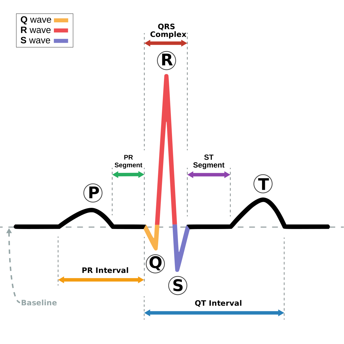
- Normal rate 60–100 bpm; <60 bradycardia, >100 tachycardia
- PR interval 120–200 ms (3–5 small boxes); >200 ms = 1st-degree AV block
- QRS <120 ms; ≥120 ms = wide (BBB, ventricular origin, hyperkalemia, pacing, WPW)
- QTc prolonged if >450 ms (men) / >470 ms (women); >500 ms = high torsades risk
- Rate shortcut: 300 ÷ large boxes between R waves
- Axis: normal if QRS upright in I and aVF; left-axis = up in I, down in aVF (confirm true LAD when lead II is also negative); right-axis = down in I, up in aVF
- STEMI: ST elevation ≥1 mm in ≥2 contiguous limb leads (or ≥2 mm in V2–V3, ≥1.5 mm in women) until proven otherwise
- New LBBB with ischemic symptoms is not an automatic STEMI — apply Sgarbossa criteria to diagnose occlusion within LBBB
- Peaked T waves = earliest sign of hyperkalemia; U waves = hypokalemia

STEMI localization by lead and artery
| ST elevation in | Wall | Culprit artery |
|---|---|---|
| V1–V4 | Anteroseptal | LAD |
| I, aVL, V5–V6 | Lateral | LCx (or diagonal) |
| II, III, aVF | Inferior | RCA (~80%) or LCx |
| ST depression V1–V3 + tall R | Posterior | RCA/LCx |
| V4R (right-sided lead) | Right ventricle | proximal RCA |
Vignette: A 58-year-old man has 45 minutes of crushing substernal chest pain, diaphoresis, and nausea. ECG shows ST elevation in II, III, and aVF (III > II) with reciprocal ST depression in I and aVL.
Diagnosis: Inferior STEMI, most often RCA occlusion (III > II and reciprocal I/aVL change favor RCA over LCx).
Next best step: Aspirin + a P2Y12 inhibitor + anticoagulation, and emergent PCI (first-medical-contact-to-device <90 min; fibrinolysis if PCI cannot be delivered within 120 min).
The trap: Inferior MI may extend to the right ventricle — obtain right-sided leads (V4R). If RV infarct is present (hypotension, clear lungs, elevated JVP), the patient is preload-dependent: give IV fluids and avoid nitrates and morphine, which drop preload and cause profound hypotension.
AV blocks — recognize and manage
| Type | ECG hallmark | Next step |
|---|---|---|
| 1st degree | PR >200 ms, constant; every P conducts | Benign — none |
| Mobitz I (Wenckebach) | PR progressively lengthens until a QRS drops | Observe; atropine if symptomatic |
| Mobitz II | Constant PR, then a sudden non-conducted P (often wide QRS) | Pacemaker — high risk of complete block |
| 3rd degree (complete) | AV dissociation — P waves and QRS fully independent | Pacemaker |
Vignette: A 72-year-old woman has fatigue, lightheadedness, and a syncopal episode. HR is 38 and regular. ECG shows P waves marching out at ~90/min and QRS complexes at ~38/min that are completely independent of one another (no fixed PR relationship).
Diagnosis: Third-degree (complete) AV block with a junctional/ventricular escape rhythm.
Next best step: Transcutaneous pacing for the unstable or symptomatic patient (atropine is usually ineffective in infranodal block), then transvenous pacing as a bridge to a permanent pacemaker. Hold AV-nodal blockers (β-blockers, non-dihydropyridine CCBs, digoxin).
Context: With an inferior MI, block is often nodal and may resolve; new complete block with an anterior MI signals extensive infarction and a worse prognosis.
The classic rhyme for second-degree AV block:
- *"Longer, longer, longer, drop — now you've got a Wenckebach"* → Mobitz I: PR progressively lengthens until a beat drops.
- *"If some P's just don't get through, then you've got a Mobitz II"* → constant PR, then a sudden non-conducted P.
Why it matters: Mobitz I is AV-nodal and benign — observe. Mobitz II is infranodal, unstable, and marches toward complete block → pacemaker.
- Delta wave + short PR + wide QRS → WPW (pre-excitation)
- Diffuse concave ST elevation + PR depression → acute pericarditis
- Electrical alternans + low voltage + tachycardia → large pericardial effusion/tamponade
- Deep symmetric T-wave inversions in V2–V3 (pain-free) → Wellens syndrome (critical proximal LAD stenosis) — do not stress test; needs catheterization
- S1Q3T3 with sinus tachycardia → PE (classic but insensitive; sinus tach is the most common finding)
- Peaked T → wide QRS → sine wave → progressive hyperkalemia; give IV calcium gluconate first to stabilize the myocardium
- Osborn (J) wave + bradycardia → hypothermia
- Coved ST elevation V1–V2 with RBBB morphology → Brugada
Practice Clinical Diagnosis now
Board-style questions, spaced-repetition flashcards, and a Socratic AI tutor — free to start.