Early Embryogenesis: Fertilization to Gastrulation
A Step 1 walkthrough of the first three weeks of human development — fertilization, the bilaminar disc, and gastrulation — mapping each germ layer to its derivatives and the classic congenital defects (neural tube defects, sacrococcygeal teratoma, caudal regression, situs inversus) that boards test by mechanism and presentation.
The First Three Weeks: A Timeline
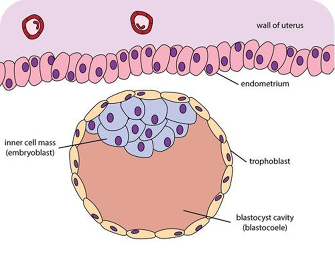
Early development is a tightly scheduled sequence, and Step 1 loves to test when and from what. Fertilization occurs in the ampulla of the uterine tube (day 0), producing a diploid zygote. Cleavage divisions yield a morula (~16 cells, day 3–4), which cavitates into a blastocyst (~day 5) composed of an outer trophoblast, an inner cell mass (embryoblast), and a fluid-filled cavity. The blastocyst hatches from the zona pellucida and implants into the endometrium around day 6–7.
The next two weeks follow a memorable rhythm. Week 2 builds the bilaminar disc (epiblast + hypoblast) — the "week of twos." Week 3 is gastrulation, when the disc becomes trilaminar (three germ layers) — the "week of threes." The single most important concept: all three germ layers derive from the epiblast as its cells migrate through the primitive streak. From these layers arise every tissue in the body, and predictable errors in their formation produce the malformations tested below.
- Fertilization happens in the ampulla; the acrosome reaction lets sperm digest through the zona pellucida (ZP3 = sperm receptor), and the cortical reaction blocks polyspermy.
- Cleavage → morula (~day 3–4) → blastocyst (~day 5) → implantation (~day 6–7).
- Week 2 = bilaminar disc (epiblast + hypoblast); the trophoblast splits into inner cytotrophoblast and outer, invasive syncytiotrophoblast.
- The syncytiotrophoblast secretes hCG, which maintains the corpus luteum (and is the basis of the pregnancy test).
- Week 3 = gastrulation: the primitive streak appears in the epiblast; all three germ layers come from epiblast.
- The notochord (from the primitive node) induces the overlying ectoderm to form the neural plate, and persists in the adult as the nucleus pulposus.
- The neural tube closes by the end of week 4 (cranial neuropore ~day 25, caudal ~day 28); failure of closure → neural tube defects.
Germ Layer → Derivatives
| Germ layer | Key derivatives (high-yield) |
|---|---|
| Surface ectoderm | Epidermis, hair, nails, sweat & mammary glands; anterior pituitary (Rathke pouch); lens & corneal epithelium; inner-ear and olfactory epithelium; tooth enamel; oral and lower-anal-canal epithelium; parotid gland |
| Neuroectoderm (neural tube) | CNS neurons and glia (astrocytes, oligodendrocytes, ependymal cells); retina & optic nerve; posterior pituitary; pineal gland |
| Neural crest | PNS (dorsal-root/autonomic ganglia, Schwann cells), adrenal medulla (chromaffin cells), melanocytes, enteric ganglia, parafollicular (C) cells, pia & arachnoid, craniofacial bones/cartilage, aorticopulmonary septum, odontoblasts |
| Mesoderm | Muscle, bone, cartilage, connective tissue, dermis; heart/vessels, blood, lymphatics, spleen; serous linings (peritoneum, pleura, pericardium); kidneys, ureters, gonads; adrenal cortex |
| Endoderm | Epithelial lining of gut & respiratory tract; liver, pancreas, gallbladder; thyroid follicular cells, parathyroids, thymus; bladder/urethra epithelium; middle-ear and auditory (Eustachian) tube lining |
Gastrulation, Neurulation, and Left–Right Asymmetry
Gastrulation begins when the primitive streak forms at the caudal epiblast. Epiblast cells invaginate through the streak: the first wave displaces the hypoblast to become definitive endoderm, the next wave becomes intraembryonic mesoderm, and the epiblast cells that remain become ectoderm. The primitive node at the cranial end of the streak gives rise to the notochord.
The notochord then induces the overlying ectoderm to undergo neurulation: the ectoderm thickens into the neural plate, elevates into neural folds, and fuses into the neural tube, while cells at the crest of the folds delaminate as neural crest. (Sonic hedgehog secreted by the notochord and floor plate later ventralizes the tube — patterning motor neurons and the floor plate.) Separately, motile cilia at the primitive node generate a leftward fluid flow that establishes the left–right body axis — when these cilia are immotile (dynein defect), laterality is randomized. Finally, the primitive streak normally regresses; if a remnant persists, it can give rise to a sacrococcygeal teratoma.

A memory scheme for pacing the early embryonic weeks. The week-2 "rule of 2s" is the well-known core; the 3s and 4s simply extend the same pattern and are worth knowing as facts even if you skip the device:
- Week 2 — "2s": 2-layer bilaminar disc (epiblast + hypoblast); trophoblast becomes 2 layers (cyto- and syncytiotrophoblast); 2 cavities form (amniotic sac + yolk sac).
- Week 3 — "3s": gastrulation makes 3 germ layers (trilaminar disc); the primitive streak appears.
- Week 4 — "4s": the heart is beating, 4 limb buds appear, and the neural tube finishes closing.
Also: "Syncytiotrophoblast Secretes hCG" — the matching first letters keep the hormone with its source.
A 26-year-old woman who took no periconceptional folate has a second-trimester screen showing markedly elevated maternal serum AFP; amniotic fluid acetylcholinesterase is positive. Ultrasound reveals a fluid-filled sac over the lumbosacral spine containing neural tissue. After birth, the infant has lower-extremity weakness and a neurogenic bladder.
Diagnosis: myelomeningocele, a neural tube defect from failure of the caudal neuropore to close (~week 4). Herniation of meninges and cord distinguishes it from a meningocele (meninges only) and from spina bifida occulta (vertebral-arch defect only, neural tube intact, normal AFP, overlying hair tuft/dimple). Teaching points: open NTDs raise AFP in both maternal serum and amniotic fluid, with amniotic acetylcholinesterase being more specific; periconceptional folate is protective, and folate antagonists (valproate, methotrexate, trimethoprim) increase risk.
Defect → Mechanism → Presentation
| Defect | Mechanism | Classic presentation |
|---|---|---|
| Anencephaly | Failure of cranial (rostral) neuropore closure | Absent forebrain & calvaria; ↑ AFP; polyhydramnios (impaired fetal swallowing) |
| Spina bifida / myelomeningocele | Failure of caudal neuropore closure | Herniated meninges ± spinal cord; motor/sensory & bladder deficits; ↑ AFP + ↑ acetylcholinesterase |
| Spina bifida occulta | Failure of vertebral-arch fusion only (neural tube intact) | Overlying hair tuft/dimple; normal AFP; usually asymptomatic |
| Sacrococcygeal teratoma | Persistent remnant of the primitive streak | Sacral mass containing all 3 germ layers; most common tumor in newborns |
| Caudal regression syndrome | Abnormal caudal mesoderm; linked to maternal diabetes | Sacral agenesis, anal atresia; severe end = lower-limb fusion (sirenomelia) |
| Situs inversus (Kartagener) | Dynein defect → immotile nodal cilia fail to set the L–R axis | Dextrocardia with bronchiectasis, sinusitis, infertility |
A term newborn is delivered with a large mass protruding from the sacrococcygeal region. Resection and histology show a mixture of tissues from all three germ layers — cartilage, gut-type glandular epithelium, and neural elements. Serum AFP is elevated and is followed as a tumor marker.
Diagnosis: sacrococcygeal teratoma, the most common tumor of the newborn, arising from a persistent remnant of the primitive streak (which normally regresses after gastrulation). Because pluripotent streak cells can give rise to all three lineages, the tumor contains ectodermal, mesodermal, and endodermal derivatives — the histologic hallmark that ties this lesion directly back to gastrulation.
- Open NTDs → ↑ maternal-serum and amniotic AFP, plus ↑ amniotic acetylcholinesterase (more specific for open NTDs).
- ↑ AFP is not specific: also seen with ventral wall defects (gastroschisis, omphalocele); low AFP suggests Down syndrome.
- Periconceptional folate reduces NTD risk; folate antagonists (valproate, methotrexate, trimethoprim) increase it.
- Maternal diabetes → caudal regression syndrome (sacral agenesis, sirenomelia) and higher congenital heart-defect risk.
- Anencephaly → polyhydramnios (the fetus cannot swallow amniotic fluid); contrast with oligohydramnios, which points to renal/GU causes.
- Persistent primitive streak → sacrococcygeal teratoma (all 3 germ layers); notochord remnants → chordoma.
Practice Embryology now
Board-style questions, spaced-repetition flashcards, and a Socratic AI tutor — free to start.