The Diaphragm & Its Openings
A board-focused walkthrough of diaphragm anatomy — origins, crura, phrenic (C3–C5) innervation and blood supply, and the T8/T10/T12 openings — anchored to the classic Step 1 clinical correlations: phrenic palsy with an elevated hemidiaphragm, Kehr-sign referred shoulder pain, and congenital versus hiatal hernias.
Overview: structure of the diaphragm
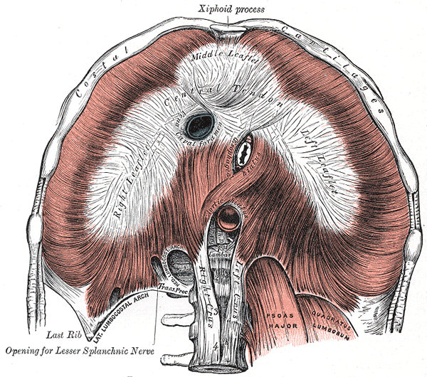
The diaphragm is the dome-shaped musculotendinous partition separating the thoracic and abdominal cavities and is the primary muscle of inspiration — when it contracts, the dome descends and flattens, increasing thoracic volume and drawing air in.
Its muscular fibers arise peripherally from three sources and converge centrally on a central tendon:
- Sternal part — two slips from the back of the xiphoid process
- Costal part — internal surfaces of the lower six ribs and costal cartilages (interdigitates with transversus abdominis)
- Lumbar part — the right and left crura plus the medial and lateral arcuate ligaments
The right crus arises from the bodies of L1–L3, the left crus from L1–L2; the right crus splits to encircle the esophageal hiatus. The central tendon fuses superiorly with the fibrous pericardium. The right dome sits higher than the left because of the underlying liver. Motor and most sensory supply come from the phrenic nerve (C3–C5).

- Motor to the ENTIRE diaphragm = phrenic nerve (C3, C4, C5) — each hemidiaphragm is driven by its ipsilateral phrenic nerve.
- Phrenic also carries sensory fibers from the central diaphragm (plus mediastinal pleura/pericardium) → irritation here refers pain to the C3–C5 shoulder tip (the anatomic basis of Kehr sign / referred diaphragmatic pain). The peripheral (costal) rim is sensory via the lower intercostal (T5–T11) and subcostal (T12) nerves.
- Blood supply: mainly the inferior phrenic arteries (off the abdominal aorta); also musculophrenic and pericardiacophrenic (from the internal thoracic) and superior phrenic (from the thoracic aorta).
- Three openings — all even numbers: T8 caval, T10 esophageal, T12 aortic.
- The right crus forms the muscular sling around the esophageal hiatus, contributing to the physiologic lower esophageal sphincter (crural "pinchcock").
- The aortic hiatus lies posterior to the diaphragm (behind the median arcuate ligament), so the aorta is not compressed as the muscle contracts.
The three openings
| Opening | Level | Part of diaphragm | Structures passing |
|---|---|---|---|
| Caval opening (vena caval foramen) | T8 | Central tendon | IVC; terminal branches of the right phrenic nerve |
| Esophageal hiatus | T10 | Muscular (right crus) | Esophagus; anterior & posterior vagal trunks; esophageal branches of the left gastric vessels |
| Aortic hiatus | T12 | Behind the diaphragm (median arcuate ligament) | Aorta; thoracic duct; azygos vein |
Levels + contents — "I ate ten eggs at twelve":
- I (IVC) at 8 (T8)
- ten (T10) eggs — (o)esophagus — plus the two vaGus (vagal) trunks
- Aorta + Thoracic duct ("AT") at 12 (T12), joined by the azygos vein
Aortic hiatus contents = "Red, White & Blue": Aorta (red), Thoracic duct (white/chyle), Azygos vein (blue).
Count the letters = the level: "vena cava" = 8 letters → T8; "aortic hiatus" = 12 letters → T12.
Phrenic roots: "C3, 4, 5 keep the diaphragm alive."
Embryologic origins — "Several Parts Build the Diaphragm": Septum transversum → central tendon, Pleuroperitoneal membranes, Body wall → muscular rim, Dorsal mesentery of esophagus → crura.
Phrenic course, development & the basis of referred pain
Each phrenic nerve forms in the neck from C3–C5 (mainly C4), descends on the surface of the anterior scalene, and enters the thorax between the subclavian artery and vein. In the mediastinum it runs anterior to the root of the lung (a key contrast with the vagus, which passes posterior to the lung root) and travels on the fibrous pericardium to reach the diaphragm.
The diaphragm develops from four embryologic parts (see mnemonic). Failure of the pleuroperitoneal membranes to close leaves a posterolateral gap → congenital diaphragmatic hernia.
Because the central diaphragm is C3–C5 in sensory origin, irritation of the overlying pleura or peritoneum (blood, pus, gallbladder disease) is referred to the shoulder tip along the C3–C4 supraclavicular dermatome — the anatomic basis for Kehr sign.
Vignette 1 — A patient with a mediastinal / hilar bronchogenic tumor develops dyspnea; CXR shows an elevated hemidiaphragm, and a fluoroscopic sniff test reveals paradoxical upward movement of that dome on inspiration. → Phrenic nerve (C3–C5) injury paralyzes the ipsilateral hemidiaphragm. Other causes: iatrogenic injury during cardiothoracic surgery (surgical traction/transection or topical-ice "cold" injury), neuralgic amyotrophy, and high cervical cord lesions. A high cervical cord lesion above the C3–C5 phrenic nucleus, or bilateral phrenic injury, abolishes diaphragmatic breathing → ventilator dependence.
Vignette 2 — After blunt abdominal trauma with splenic rupture, a hypotensive patient complains of left shoulder pain (Kehr sign). → Blood irritates the central diaphragmatic peritoneum, whose C3–C4 afferents refer pain to the supraclavicular shoulder dermatome. The same mechanism explains shoulder-tip pain from a subphrenic abscess or, on the right, gallbladder disease.
Newborn with respiratory distress, a scaphoid abdomen, bowel sounds in the left chest, and the mediastinum shifted to the right → congenital diaphragmatic hernia (Bochdalek type): a posterolateral defect from failed closure of the pleuroperitoneal membrane, usually left-sided. Herniated abdominal viscera compress the developing lung → pulmonary hypoplasia, the chief cause of mortality.
Older adult with heartburn and regurgitation → sliding hiatal hernia (~95% of hiatal hernias): the GE junction and cardia slide upward through the esophageal hiatus into the posterior mediastinum; strongly associated with GERD.
Paraesophageal (rolling) hernia: the gastric fundus herniates alongside the esophagus while the GE junction stays below the diaphragm; reflux is less prominent, but there is a real risk of incarceration/strangulation.
Morgagni hernia: an anterior/retrosternal defect (foramen of Morgagni), rare, more often right-sided, and usually asymptomatic/incidental in adults.
Hernia comparison
| Hernia | Defect / location | Typical side | Classic clue |
|---|---|---|---|
| Bochdalek | Posterolateral (pleuroperitoneal membrane) | Left | Neonatal distress + pulmonary hypoplasia; "Back, Big, Left" |
| Morgagni | Anterior / retrosternal (foramen of Morgagni) | Right | Rare, often incidental in adults; "anteri-Mor" |
| Sliding hiatal | GE junction slides through esophageal hiatus | Midline | GERD; most common (~95%) |
| Paraesophageal | Fundus beside esophagus, GE junction fixed | Midline | Reflux mild, but strangulation risk |
Practice Anatomy now
Board-style questions, spaced-repetition flashcards, and a Socratic AI tutor — free to start.