Dementia & Neurodegenerative Disease
A boards-focused neurology lesson on dementia and neurodegenerative disease, moving from proteinopathy pathophysiology through pattern-based localization, comparison of the major dementias (Alzheimer, vascular, Lewy body, frontotemporal, NPH, prion), and the classic next-best-step decisions tested on Step 1 and Step 2 CK.
Overview & Pathophysiology
Dementia (major neurocognitive disorder) is an acquired, progressive decline in ≥1 cognitive domain (memory, language, executive function, visuospatial skill, behavior) severe enough to impair daily function, with a normal level of consciousness — distinguishing it from delirium. Most neurodegenerative dementias are proteinopathies, defined by a misfolded protein:
- Alzheimer — extracellular amyloid-β (Aβ) plaques + intracellular tau tangles
- Lewy body / Parkinson — α-synuclein
- Frontotemporal — tau or TDP-43
- Prion (CJD) — misfolded PrP^sc
Vascular dementia results from ischemic injury — preventable/modifiable by controlling vascular risk factors, though established infarcts are not reversible. Normal pressure hydrocephalus (NPH), from disordered CSF dynamics, is the classic potentially reversible neurodegenerative-mimicking dementia (with shunting). Boards test the pattern of onset (insidious vs stepwise vs rapid) and the first cognitive domain affected to localize the diagnosis — and always screen for the truly reversible mimics (B12, thyroid, neurosyphilis) before attributing decline to a neurodegenerative process.
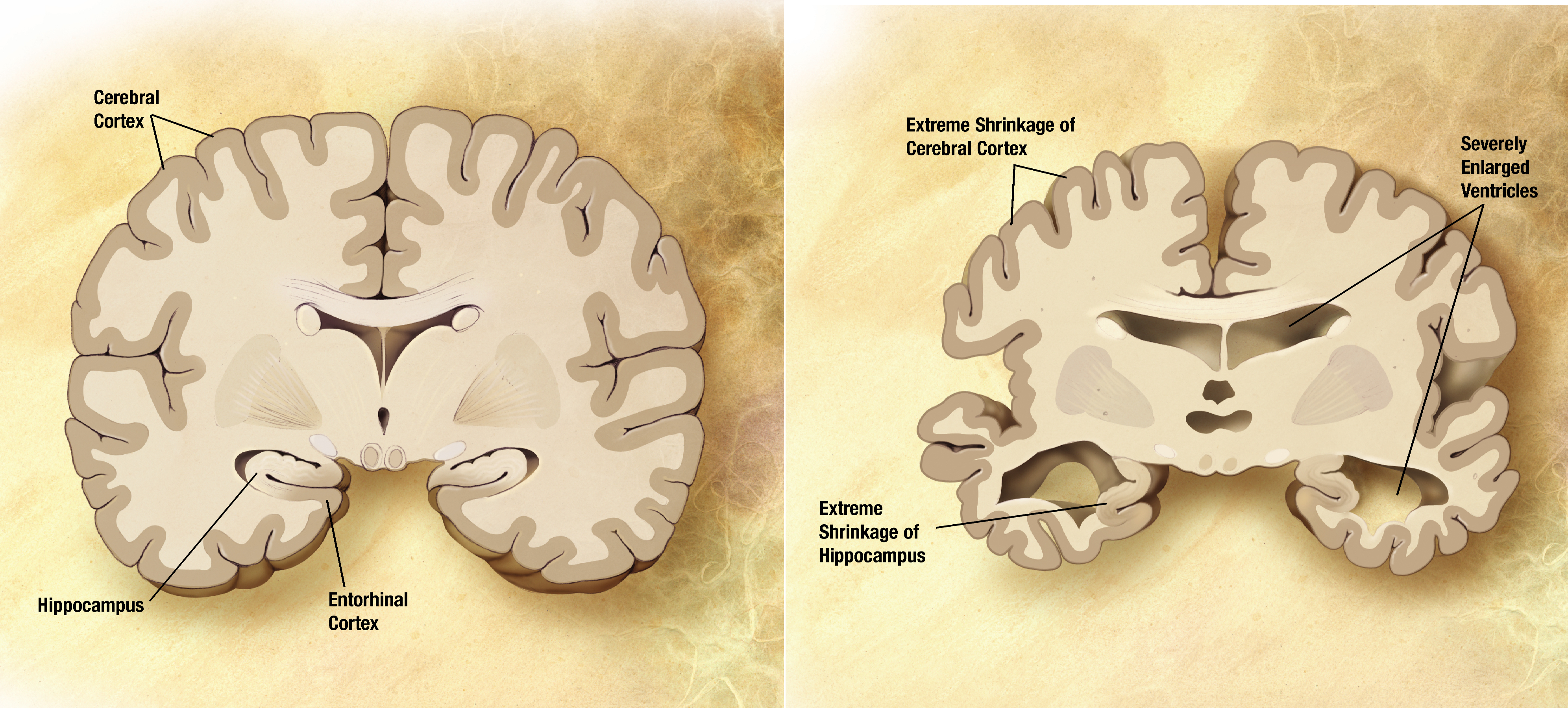
- Alzheimer: insidious anterograde memory loss; getting lost, word-finding trouble; ↓ACh (nucleus basalis of Meynert)
- Vascular: stepwise decline + focal deficits; HTN/stroke/AF history
- Lewy body (DLB): fluctuating cognition, recurrent visual hallucinations, parkinsonism, REM sleep behavior disorder, neuroleptic sensitivity
- Frontotemporal (Pick): young (45–65), early personality/behavior change or aphasia, memory spared early
- NPH: wet, wacky, wobbly (incontinence, dementia, magnetic gait)
- Prion (CJD): rapidly progressive dementia (wks–mos) + startle myoclonus
- Huntington: AD, CAG repeat (HTT, chr 4), chorea + subcortical dementia, caudate atrophy
- Reversible mimics: B12 deficiency, hypothyroidism, neurosyphilis, chronic subdural, depression (pseudodementia)
Comparison of Major Dementias
| Disease | Classic clues | Pathology | Key test finding |
|---|---|---|---|
| Alzheimer | Insidious anterograde memory loss, visuospatial/word-finding decline | Aβ plaques + tau tangles; ↓ACh | ↓CSF Aβ42, ↑p-tau; hippocampal atrophy; amyloid PET+ |
| Vascular | Stepwise decline, focal signs, HTN/stroke hx | Multi-infarct / small-vessel ischemia | MRI: cortical + subcortical infarcts, white-matter hyperintensities |
| Lewy body | Fluctuating cognition, visual hallucinations, parkinsonism, REM behavior disorder | α-synuclein Lewy bodies | Severe neuroleptic sensitivity; ↓DAT uptake (DaTscan SPECT) |
| Frontotemporal | Young; behavior/personality or aphasia; memory spared early | Tau (Pick bodies) or TDP-43 | MRI: focal fronto-temporal (knife-edge) atrophy |
| NPH | Wet, wacky, wobbly | Impaired CSF resorption (communicating) | Ventriculomegaly out of proportion to atrophy; LP tap test |
| Prion (CJD) | Rapid dementia + myoclonus | PrP^sc spongiform change | DWI cortical ribboning + basal ganglia; EEG PSWCs; CSF RT-QuIC/14-3-3 |
A 72-year-old man has 1 year of fluctuating confusion, recurrent well-formed visual hallucinations of small animals, and mild rigidity/bradykinesia. His wife reports he acts out dreams and has fallen twice. In the ED he received haloperidol and became severely rigid, febrile, and unresponsive.
- Diagnosis: Dementia with Lewy bodies (α-synuclein). Dementia arising before, with, or within 1 year of parkinsonism = DLB (vs Parkinson disease dementia, where dementia follows established motor disease by >1 year). The catastrophic reaction reflects classic neuroleptic sensitivity.
- Next best step: Stop the antipsychotic; avoid typical/atypical neuroleptics (especially haloperidol). Treat cognition with a cholinesterase inhibitor (rivastigmine/donepezil); if psychosis is dangerous, use low-dose quetiapine or pimavanserin cautiously. Treat REM sleep behavior disorder with melatonin.
- NPH triad — "Wet, Wacky, Wobbly": urinary incontinence, dementia, magnetic/apraxic gait. Gait responds best to shunting.
- Alzheimer cortical deficits — the 4 A's: Amnesia, Aphasia, Apraxia, Agnosia.
- Early-onset familial AD & Down syndrome — think chromosome 21: APP gene (chr 21); trisomy 21 → early Alzheimer. (Other familial genes: PSEN1/chr 14, PSEN2/chr 1.)
Alzheimer Disease — Deep Dive
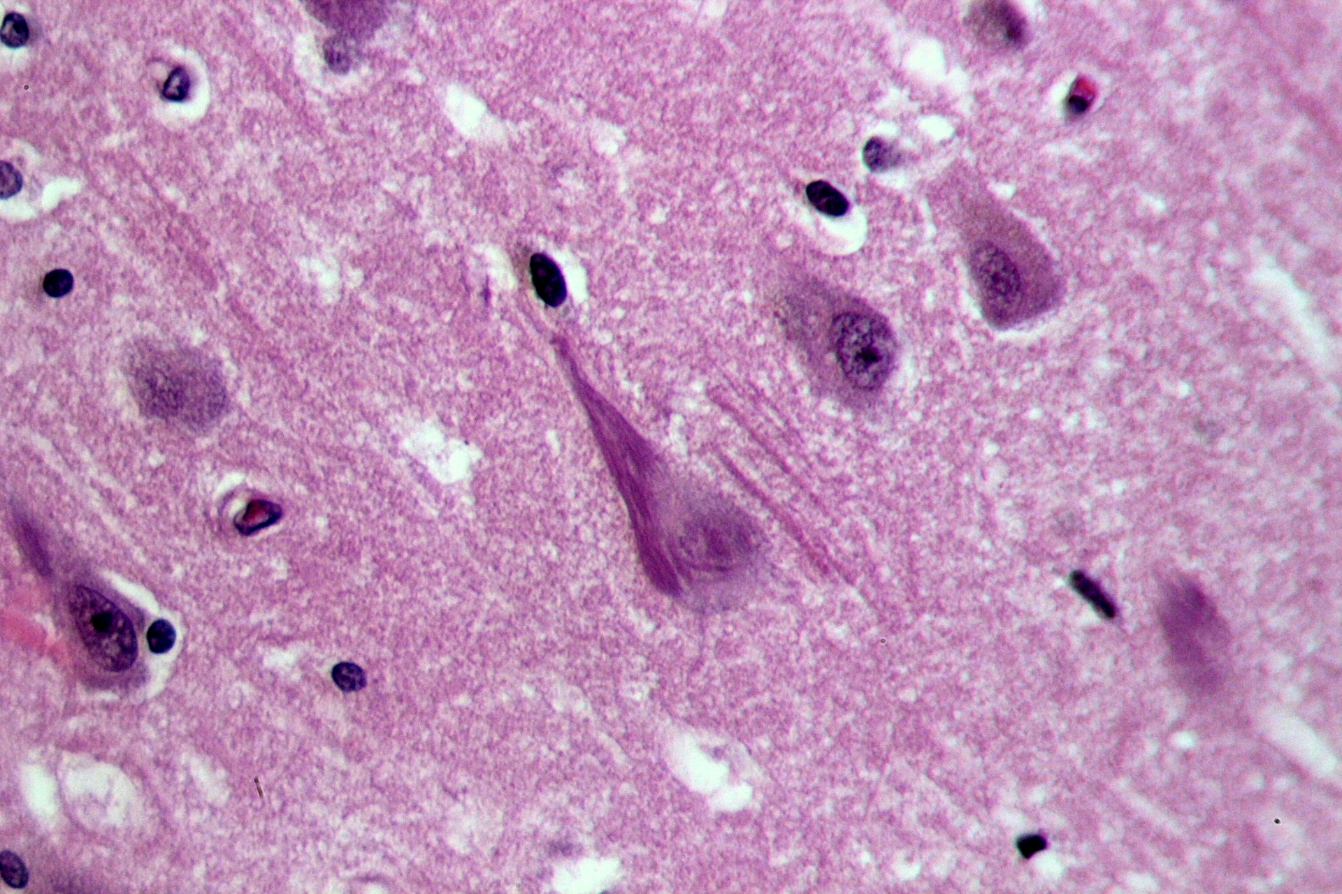
Alzheimer disease is the most common dementia. Amyloid precursor protein (APP) is cleaved by β- and γ-secretase into Aβ42, which aggregates into extracellular neuritic (senile) plaques; intracellular neurofibrillary tangles are paired helical filaments of hyperphosphorylated tau. Loss of cholinergic neurons in the nucleus basalis of Meynert lowers ACh. Gross: diffuse cortical atrophy (esp. hippocampus/temporal) with hydrocephalus ex vacuo.
Genetics: late-onset risk rises with APOE ε4 (ε4/ε4 highest; ε2 protective); autosomal-dominant early-onset from APP, PSEN1, PSEN2; Down syndrome → early AD.
Biomarkers: CSF ↓Aβ42, ↑total/phospho-tau; amyloid PET; emerging plasma p-tau217.
Treatment: cholinesterase inhibitors (donepezil, rivastigmine, galantamine); add memantine (NMDA antagonist) in moderate–severe disease. Anti-amyloid monoclonals lecanemab / donanemab modestly slow early AD but cause ARIA (edema/microhemorrhage), highest in APOE ε4 homozygotes — require amyloid confirmation + serial MRI monitoring.
A 74-year-old woman has 6 months of a progressive shuffling, magnetic gait ("feet stuck to the floor"), worsening short-term memory, and new urinary incontinence. Neuro exam shows no focal weakness. Non-contrast CT shows ventricular enlargement out of proportion to cortical atrophy (elevated Evans index).
- Diagnosis: Normal pressure hydrocephalus — a communicating, potentially reversible hydrocephalus with normal opening pressure.
- Next best step: Large-volume lumbar puncture ("tap test") — remove ~30–50 mL CSF and reassess gait; transient improvement predicts response to a ventriculoperitoneal (VP) shunt.
- Pearl: Gait improves most with shunting; dementia improves least — so evaluate early. Always exclude reversible mimics (B12, TSH, RPR) before attributing decline to a neurodegenerative process.
- Delirium vs dementia: delirium is acute, fluctuating, with impaired attention/consciousness and often reversible — rule out and treat first.
- Always exclude reversible/secondary causes: check B12, TSH, CMP, CBC; consider RPR/FTA (neurosyphilis) and HIV; obtain MRI/CT for tumor, chronic subdural, NPH, or vascular disease.
- Pseudodementia = depression mimicking dementia (poor effort, frequent "I don't know") — treat the depression.
- Suspected NPH → large-volume LP (tap test) before shunting.
- DLB → avoid antipsychotics (severe neuroleptic sensitivity).
- Rapidly progressive dementia + myoclonus → think CJD: MRI DWI, EEG, CSF RT-QuIC (most specific).
- Screening: MMSE/MoCA; definitive AD or prion diagnosis relies on biomarkers or autopsy.
Practice Neuro now
Board-style questions, spaced-repetition flashcards, and a Socratic AI tutor — free to start.