Cystic Kidney Disease (ADPKD, ARPKD)
A boards-focused walk through ADPKD and ARPKD — from the polycystin/fibrocystin ciliopathy mechanism to presentation, ultrasound-based diagnosis (Pei–Ravine criteria), classic extrarenal traps, and management including ACE inhibitors and tolvaptan. Built around vignette buzzwords and next-best-step decisions the way the boards test them.
Pathophysiology — a ciliopathy of the tubule
Cystic kidney disease is a spectrum of ciliopathies in which fluid-filled cysts bud from renal tubular epithelium. The two board-classic hereditary forms are ADPKD (adults) and ARPKD (infants).
In ADPKD, mutations in PKD1 (chromosome 16, polycystin-1, ~85%, more severe) or PKD2 (chromosome 4, polycystin-2, ~15%, milder) disrupt polycystins in the primary cilium. Loss of ciliary mechanosensation raises intracellular cAMP, driving epithelial proliferation and fluid secretion → progressive bilateral cysts that destroy nephrons over decades. Cysts arise from any nephron segment, and a somatic 'second hit' is needed for an individual cyst to form.
In ARPKD, mutation of PKHD1 (chromosome 6) impairs fibrocystin, producing fusiform dilation of collecting ducts plus congenital hepatic fibrosis.
Expanding cysts cause mass effect and RAAS activation → hypertension. Because nephrons are lost gradually, ADPKD is usually silent until adulthood, while ARPKD declares itself at (or before) birth.
- Most common inherited kidney disease (~1:400–1000); autosomal dominant, near-complete penetrance
- PKD1 (chr 16) ≈ 85%, ESRD ~50s; PKD2 (chr 4) milder, ESRD ~70s
- Hypertension is often the earliest sign (RAAS activation)
- Flank pain, painless gross hematuria (cyst rupture), nephrolithiasis, recurrent UTIs / cyst infection
- Bilateral enlarged, palpable kidneys
- Extrarenal: berry (saccular) aneurysm → subarachnoid hemorrhage; hepatic cysts (most common extrarenal manifestation, worse in women/estrogen); mitral valve prolapse; thoracic aortic aneurysm/dissection; colonic diverticulosis
- Leading cause of death = cardiovascular (not SAH)
- Urinalysis: hematuria, mild proteinuria (usually <1 g/day), early urine-concentrating defect (nocturia)
- Autosomal recessive; PKHD1 (chr 6) → defective fibrocystin; presents in neonates/infants
- Bilaterally enlarged, smooth, echogenic kidneys on ultrasound with poor corticomedullary differentiation (dilated collecting ducts)
- In utero: poor urine output → oligohydramnios → Potter sequence (pulmonary hypoplasia, limb/facial deformities) → neonatal respiratory failure = main early killer
- Congenital hepatic fibrosis → portal hypertension, hepatosplenomegaly, esophageal varices; associated Caroli disease (intrahepatic bile-duct dilation)
- Systemic hypertension and progressive CKD in survivors
- Hepatic involvement is the rule, not the exception
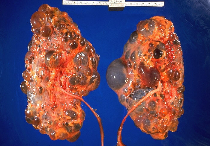
ADPKD vs ARPKD
| Feature | ADPKD | ARPKD |
|---|---|---|
| Gene (chromosome) | PKD1 (16), PKD2 (4) | PKHD1 (6) |
| Protein | Polycystin-1 / -2 | Fibrocystin |
| Onset | Adulthood (30s–40s) | Neonatal / infancy |
| Kidneys | Large; cysts in cortex and medulla | Large, smooth, echogenic; dilated collecting ducts |
| Liver | Hepatic cysts | Congenital hepatic fibrosis, Caroli |
| Vascular / cardiac | Berry aneurysm, MVP, aortic dissection | — |
| Perinatal | — | Oligohydramnios → Potter, pulmonary hypoplasia |
| Course | ESRD ~50s–70s | High neonatal mortality; childhood ESRD |
A 35-year-old man has newly diagnosed hypertension and an episode of painless gross hematuria. His father died of a 'brain bleed.' Exam: bilateral palpable flank masses; creatinine mildly elevated.
- Diagnosis: ADPKD
- Best initial / confirmatory test: renal ultrasound — age-adjusted Pei–Ravine criteria in an at-risk (family-history-positive) person: age 15–39 → ≥3 cysts total; 40–59 → ≥2 per kidney; ≥60 → ≥4 per kidney
- Next best step given family history of aneurysm/SAH: screen brain with MR angiography (indicated with family history of aneurysm/SAH or high-risk occupation — not routine for everyone)
- If he presents with 'worst headache of life': non-contrast head CT for subarachnoid hemorrhage
- First-line antihypertensive: ACE inhibitor / ARB
A neonate born after a pregnancy complicated by oligohydramnios has respiratory distress, low-set ears, a flattened nose, limb contractures, and bilateral abdominal masses. Ultrasound: massively enlarged, echogenic kidneys.
- Diagnosis: ARPKD with Potter sequence
- Immediate threat to life: pulmonary hypoplasia → respiratory failure (not renal failure)
- Next best step: stabilize the airway / respiratory support first, then confirm with renal + hepatic ultrasound
- Anticipate long-term: congenital hepatic fibrosis → portal hypertension / variceal bleeding; systemic hypertension
- Buzzword: oligohydramnios + bilateral flank masses in a neonate = ARPKD

POTTER — the downstream consequences of oligohydramnios:
- P — Pulmonary hypoplasia (the lethal one)
- O — Oligohydramnios (the trigger)
- T — Twisted face (Potter facies: low-set ears, flat nose, recessed chin)
- T — Twisted skin (redundant, compressed fetal skin)
- E — Extremity deformities (clubfeet, limb contractures)
- R — Renal cause of the low fluid (ARPKD, bilateral renal agenesis, posterior urethral valves)
For ADPKD, anchor the berry aneurysm to its rupture: the 'worst headache of life' = subarachnoid hemorrhage.
Don't confuse these cystic diseases
| Disease | Inheritance | Classic clues |
|---|---|---|
| ADPKD | AD (PKD1/2) | HTN, hematuria, berry aneurysm, hepatic cysts; large kidneys |
| ARPKD | AR (PKHD1) | Neonate, Potter, hepatic fibrosis; large kidneys |
| ADTKD / medullary cystic | AD (UMOD, MUC1) | Bland urine, small kidneys, gout, ESRD in adulthood |
| Medullary sponge kidney | Sporadic | Recurrent Ca stones/UTIs, hematuria; usually benign |
| Acquired cystic disease | Acquired (dialysis) | Long-term dialysis → ↑ renal cell carcinoma |
| Simple renal cyst | Acquired | Incidental, benign (Bosniak I) |
- BP control, first-line: ACE inhibitor / ARB (RAAS-driven HTN); rigorous control slows kidney-volume growth (HALT-PKD)
- Tolvaptan (vasopressin V2-receptor antagonist, lowers cAMP) slows eGFR decline in rapidly progressing ADPKD — monitor LFTs (hepatotoxicity); expect aquaresis/thirst
- High fluid intake (suppresses ADH), low sodium
- Cyst infection: use lipophilic antibiotics that penetrate cysts (fluoroquinolone, TMP-SMX)
- ESRD → dialysis or kidney transplant (curative for the renal disease)
- Screen for intracranial aneurysm with MRA only if family history of aneurysm/SAH or high-risk occupation
- Offer genetic counseling; screen at-risk first-degree relatives by ultrasound
Practice Renal now
Board-style questions, spaced-repetition flashcards, and a Socratic AI tutor — free to start.