Community-Acquired Pneumonia
A boards-focused walkthrough of community-acquired pneumonia: the CXR-confirmed diagnosis, typical vs. atypical organism buzzwords, CURB-65 disposition, and the 2019 ATS/IDSA empiric antibiotic tiers — all anchored to the "next best step" the exam rewards.
Framing CAP
Community-acquired pneumonia (CAP) is an acute infection of the lung parenchyma acquired outside the hospital (or within <48 h of admission). *Streptococcus pneumoniae* remains the single most common bacterial cause.
- "Typical" pathogens (pneumococcus, Haemophilus influenzae, Moraxella catarrhalis) → lobar consolidation with productive cough and high fever.
- "Atypical" pathogens (Mycoplasma pneumoniae, Chlamydophila pneumoniae, Legionella) → often more indolent with a diffuse/interstitial picture and prominent extrapulmonary symptoms — but Legionella is the exception that frequently causes severe pneumonia.
- Respiratory viruses (influenza, RSV, SARS-CoV-2) are increasingly recognized.
Classic exam: fever, productive cough, pleuritic chest pain, dyspnea, with focal crackles, bronchial breath sounds, egophony (E→A), dullness to percussion, and increased tactile fremitus. Diagnosis requires a compatible clinical picture PLUS a new infiltrate on chest radiograph. The 2019 ATS/IDSA guideline formally retired the term "HCAP."

- A chest X-ray is required to diagnose CAP — a new infiltrate/consolidation confirms it; a truly normal CXR argues against it.
- *S. pneumoniae* = most common bacterial cause overall.
- CURB-65 (or the more detailed PSI) drives disposition: home vs. ward vs. ICU.
- Outpatient, previously healthy: high-dose amoxicillin or doxycycline; a macrolide alone only where local pneumococcal resistance <25%.
- Outpatient w/ comorbidities or inpatient (non-severe): β-lactam + macrolide OR a respiratory fluoroquinolone (levofloxacin, moxifloxacin).
- ICU/severe: β-lactam + macrolide OR β-lactam + fluoroquinolone — never fluoroquinolone monotherapy in the ICU.
- Blood + sputum cultures and Legionella/pneumococcal urinary antigens are for severe/inpatient disease — not routine for healthy outpatients.
- Add MRSA (vancomycin or linezolid) or *Pseudomonas* coverage only with prior isolation of the organism or validated local risk factors — and obtain cultures.
- Post-influenza pneumonia → think *S. aureus (incl. MRSA) and S. pneumoniae*.
- Duration ≥5 days, continued until afebrile 48–72 h and clinically stable.
Organism Buzzwords (typical vs. atypical)
| Vignette buzzword | Likely organism | Pearl / next test |
|---|---|---|
| Rust-colored sputum, lobar consolidation | Streptococcus pneumoniae | Most common; pneumococcal urine antigen |
| Currant-jelly sputum, alcoholic/aspiration, cavitary upper lobe | Klebsiella pneumoniae | "Bulging fissure" on CXR |
| College student, "walking pneumonia," bullous myringitis | Mycoplasma pneumoniae | Cold agglutinins → cold AIHA |
| High fever, diarrhea, hyponatremia, ↑LFTs, water/AC/cruise exposure | Legionella pneumophila | Urine antigen; grows on buffered charcoal yeast extract |
| Post-influenza, necrotizing/cavitary, empyema | Staphylococcus aureus | Cover MRSA if risk factors |
| Bird/parrot exposure | Chlamydophila psittaci | Occupational/pet history |
| Farm animals / parturient cats (Q fever) | Coxiella burnetii | — |
| COPD exacerbation with pneumonia | H. influenzae / M. catarrhalis | — |
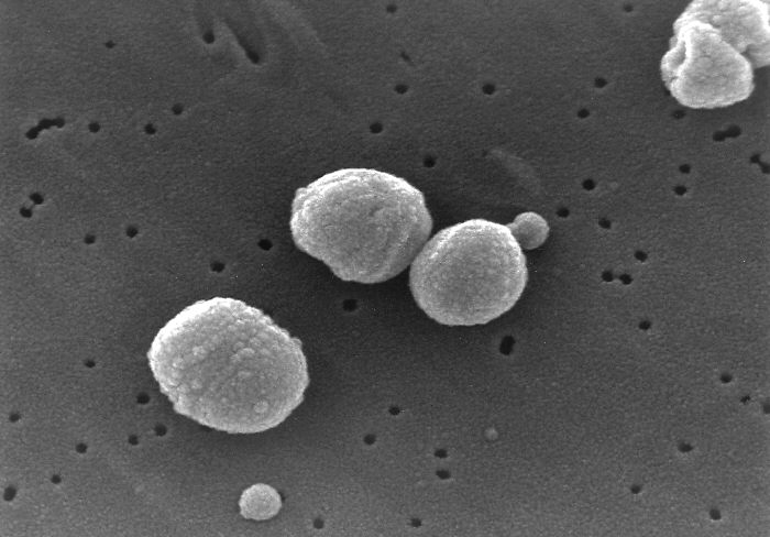
Vignette 1 — "Walking pneumonia." A 22-year-old college student has 10 days of malaise, low-grade fever, and a dry hacking cough. He "looks well," but CXR shows patchy bilateral interstitial infiltrates out of proportion to exam. CBC shows mild anemia; cold-agglutinin test is positive.
- Dx: Mycoplasma pneumoniae with cold-agglutinin hemolytic anemia.
- NEXT BEST STEP: outpatient azithromycin (macrolide) or doxycycline. β-lactams are useless — Mycoplasma has no cell wall.
Vignette 2 — Legionnaires'. A 60-year-old smoker returns from a cruise with high fever, watery diarrhea, confusion, and labs showing Na 128 and elevated transaminases; CXR shows patchy consolidation.
- NEXT BEST STEP: urine Legionella antigen; treat with a respiratory fluoroquinolone or macrolide (β-lactams fail — intracellular organism).
The Workup Ladder & Severity
Boards reward a predictable sequence once CAP is suspected:
- Confirm with a chest X-ray (new infiltrate).
- Assess severity with CURB-65 to decide disposition.
- Obtain cultures + urinary antigens only if inpatient/severe.
- Start empiric antibiotics promptly — do not delay treatment waiting for cultures.
Severe CAP (IDSA: ≥1 major criterion — septic shock needing vasopressors, or respiratory failure requiring mechanical ventilation; or ≥3 minor criteria) → ICU.
A pneumonia that fails to improve or clear on repeat imaging → think parapneumonic effusion/empyema, a resistant or unusual organism, an obstructing malignancy, or a non-infectious mimic (e.g., organizing pneumonia, PE) → CT chest ± bronchoscopy.
One point each — predicts 30-day mortality and where the patient goes:
- C — Confusion (new)
- U — Urea / BUN >19 mg/dL (>7 mmol/L)
- R — Respiratory rate ≥30/min
- B — Blood pressure: SBP <90 or DBP ≤60
- 65 — Age ≥65 years
Score 0–1 → home · 2 → ward admission · ≥3 → admit, evaluate for ICU. (CRB-65, dropping urea, works when labs are unavailable.)
Empiric Antibiotics by Setting (2019 ATS/IDSA)
| Setting | Preferred empiric regimen |
|---|---|
| Outpatient, no comorbidities | Amoxicillin 1 g PO TID OR doxycycline OR macrolide (only if pneumococcal resistance <25%) |
| Outpatient with comorbidities\* | (β-lactam: amox-clav or cefpodoxime) + (macrolide or doxycycline) OR respiratory fluoroquinolone (levofloxacin 750 mg / moxifloxacin) |
| Inpatient, non-severe | (β-lactam: ceftriaxone / cefotaxime / ampicillin-sulbactam) + macrolide OR respiratory fluoroquinolone monotherapy |
| Inpatient, severe (ICU) | β-lactam + macrolide OR β-lactam + fluoroquinolone — no FQ monotherapy |
| MRSA risk / prior MRSA | Add vancomycin or linezolid (send cultures) |
| *Pseudomonas* risk / prior isolate | Antipseudomonal β-lactam (pip-tazo, cefepime, meropenem) |
A 58-year-old admitted for CAP is still febrile on day 3 of ceftriaxone + azithromycin. Repeat exam: dullness and absent breath sounds at the left base; CXR shows a moderate layering pleural effusion.
- NEXT BEST STEP: diagnostic thoracentesis (with decubitus film/ultrasound to confirm free-flowing fluid).
- Fluid indicating a complicated parapneumonic effusion / empyema — pH <7.20, glucose <60 mg/dL, positive Gram stain/culture, or frank pus — mandates chest-tube (tube thoracostomy) drainage, not antibiotics alone.
- Loculated/organized collections → intrapleural tPA + DNase or VATS decortication.
Practice Internal Medicine now
Board-style questions, spaced-repetition flashcards, and a Socratic AI tutor — free to start.