Colorectal Surgical Emergencies (Perforation, Volvulus, Ischemia)
A Step 2 CK-focused lesson on the three colonic surgical emergencies — perforation, volvulus, and ischemic colitis — built around board buzzwords and next-best-step decisions (who gets decompressed, who gets antibiotics, and who goes to the OR).
The Big Picture
Colorectal surgical emergencies share a final common pathway: a distended, obstructed, or ischemic colon that can progress to transmural necrosis, perforation, and feculent peritonitis. On the boards the task is pattern recognition plus the next-best-step — decide who gets endoscopic decompression, who gets IV fluids and antibiotics, and who goes straight to the OR.
Three archetypes dominate Step 2 CK:
- Perforation — usually complicating diverticulitis or an obstructing colon cancer.
- Volvulus — sigmoid (far more common) and cecal.
- Ischemic colitis — a low-flow watershed injury of the colon.
The unifying red flags that mandate laparotomy — regardless of cause — are peritonitis, hemodynamic instability, free intraperitoneal air, and bowel gangrene. Everything else is about matching the vignette to the least-invasive correct move.
- Diverticulitis is the most common cause of colonic perforation; obstructing colon cancer and iatrogenic colonoscopy injury are next.
- CT abdomen/pelvis with IV contrast is the diagnostic test of choice for diverticulitis and suspected perforation.
- Free air under the diaphragm on upright CXR (or extraluminal air on CT) = perforated viscus → surgery.
- Hinchey: I = pericolic abscess, II = pelvic/distant abscess, III = purulent peritonitis, IV = feculent peritonitis.
- Abscess >3–4 cm → CT-guided percutaneous drainage + IV antibiotics; interval elective resection.
- Hinchey III–IV (diffuse peritonitis) → emergent Hartmann procedure = sigmoid resection + end colostomy (no anastomosis in a contaminated/unstable field).
- Uncomplicated diverticulitis → bowel rest ± antibiotics; colonoscopy 6–8 weeks later to exclude cancer.
A 68-year-old man with prior diverticulitis has 2 days of worsening LLQ pain, now diffuse. Temp 39°C, HR 120, BP 88/54. The abdomen is rigid with rebound. Upright CXR shows free air under both hemidiaphragms.
- Diagnosis: perforated diverticulitis with purulent/feculent peritonitis (Hinchey III–IV).
- Next best step: IV fluids + broad-spectrum antibiotics, then emergent laparotomy with Hartmann procedure.
Trap: Do NOT choose outpatient antibiotics, percutaneous drainage, or delayed colonoscopy here. Free air + peritonitis + shock = OR. Percutaneous drainage is only for a contained abscess in a stable patient without diffuse peritonitis.
An 80-year-old nursing-home resident with chronic constipation has 3 days of massive abdominal distension, obstipation, and cramping. The abdomen is tympanitic and non-tender. Plain film shows a hugely dilated "coffee-bean" loop arising from the pelvis and pointing toward the RUQ.
- Diagnosis: sigmoid volvulus.
- Next best step (no peritonitis/ischemia): endoscopic detorsion via flexible sigmoidoscopy with rectal tube placement — the classic "decompress, don't cut yet" answer. Endoscopy also assesses viability: dark, non-viable mucosa or blood signals ischemia.
- Then: elective sigmoid colectomy during the same admission — recurrence after detorsion alone is high (~40–60%).
- If peritonitis, gangrene, or failed detorsion → emergent sigmoidectomy (Hartmann).
Sigmoid vs. Cecal Volvulus
| Feature | Sigmoid volvulus | Cecal volvulus |
|---|---|---|
| Frequency | Most common (~60–75%) | Less common (~25–40%) |
| Typical patient | Elderly, bedridden, chronic constipation, psych/neuro meds | Younger adults; congenital mobile cecum, pregnancy |
| "Coffee-bean" apex points to | RUQ (arises from LLQ) | LUQ/epigastrium (arises from RLQ) |
| First-line treatment | Endoscopic detorsion + rectal tube, then elective sigmoid colectomy | Surgery — right hemicolectomy (detorsion usually fails) |
| If ischemia/peritonitis | Emergent resection (Hartmann) | Emergent resection |
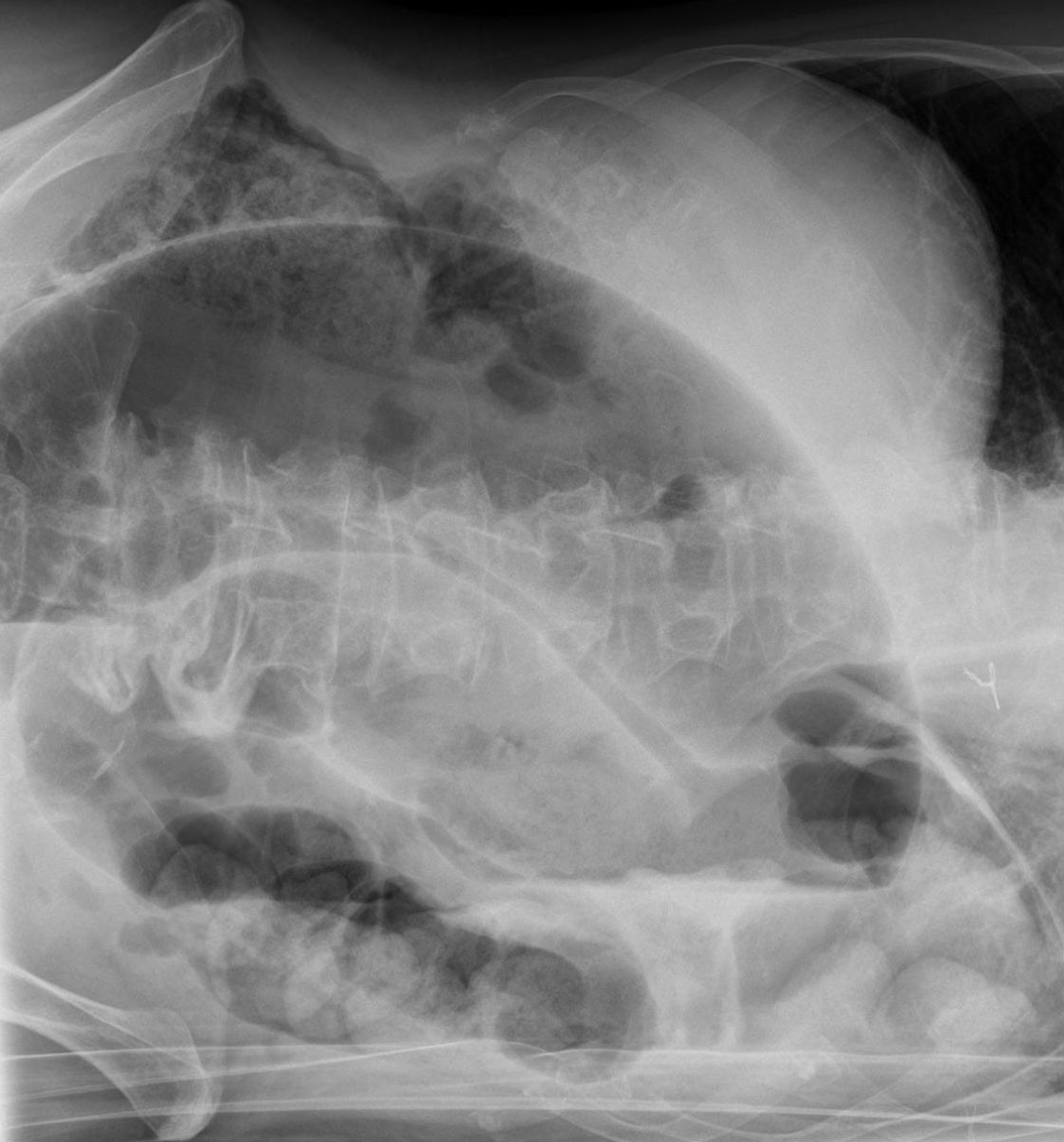
A 72-year-old woman develops sudden crampy LLQ pain followed within a day by bloody diarrhea, shortly after open AAA repair (or an episode of hypotension/hypovolemia). She is only mildly tender in the LLQ; lactate is near-normal.
- Diagnosis: ischemic colitis — a low-flow watershed injury (classically splenic flexure and rectosigmoid).
- Diagnosis clues: CT shows "thumbprinting" (submucosal edema/hemorrhage) and segmental wall thickening; colonoscopy is the most sensitive test and shows segmental mucosal ischemia that spares the rectum (dual blood supply).
- Management: mostly supportive — IV fluids, bowel rest, and antibiotics; most non-gangrenous cases resolve. Surgery only for gangrene, perforation, fulminant colitis, or a later stricture.
Contrast: pain out of proportion to exam points instead to acute mesenteric ischemia (small bowel, SMA).
Ischemic Colitis vs. Acute Mesenteric Ischemia
| Feature | Ischemic colitis | Acute mesenteric ischemia |
|---|---|---|
| Territory | Watershed colon (SMA–IMA border) | SMA — small bowel |
| Mechanism | Low flow / hypotension | Embolus (AFib), thrombosis, NOMI, venous |
| Pain | Mild, LLQ, + bloody diarrhea | Pain out of proportion to exam |
| Lactate/acidosis | Usually normal early | Elevated (marks necrosis) |
| Best test | Colonoscopy; CT "thumbprinting" | CT angiography |
| Treatment | Supportive | Revascularization + resect necrotic bowel |
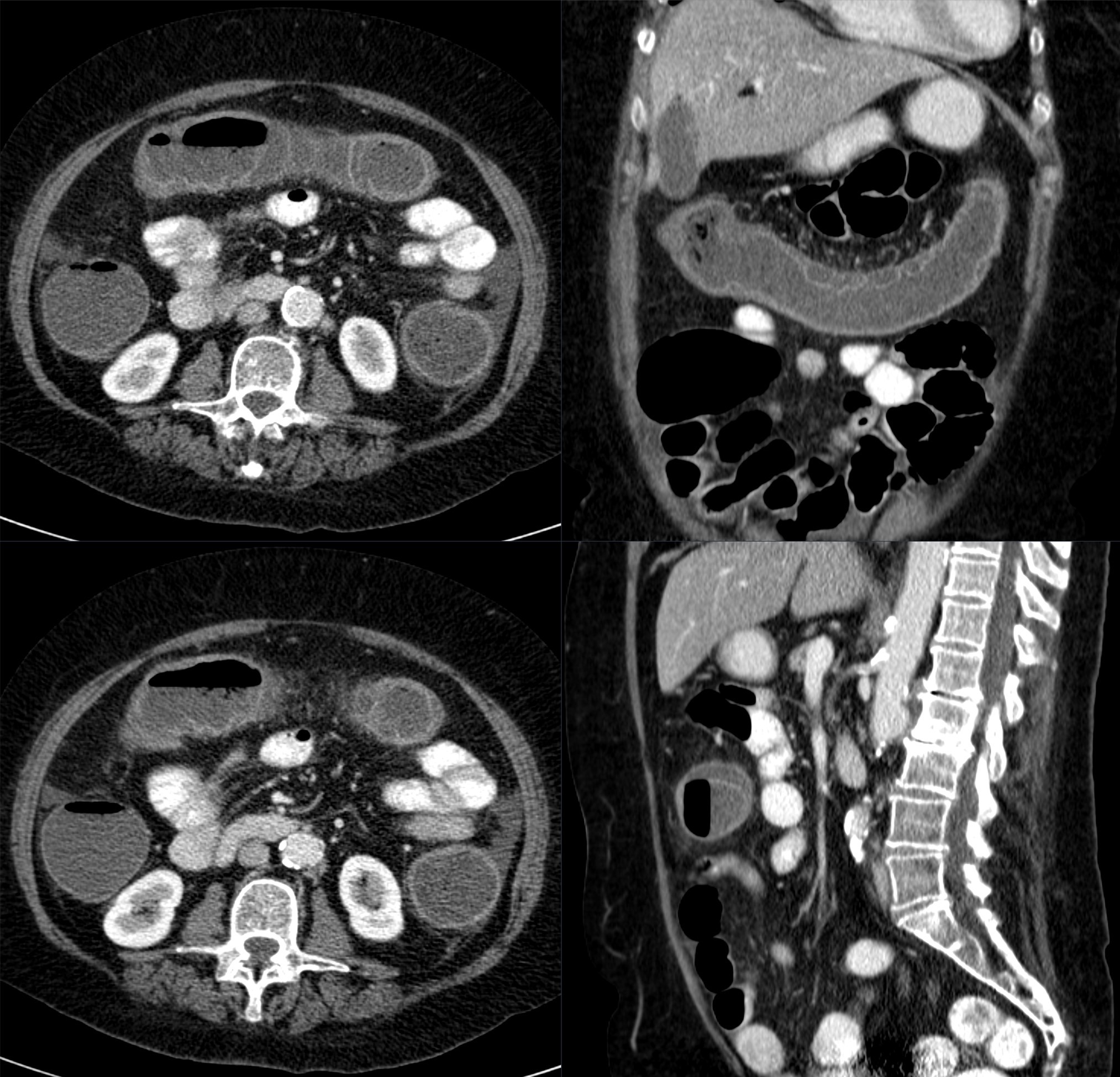
- Peritonitis, hemodynamic instability, free air, or gangrene → OR, regardless of the underlying cause.
- Sigmoid volvulus without ischemia → endoscopic detorsion first, then elective colectomy same admission.
- Cecal volvulus → primary surgery (right hemicolectomy) — detorsion usually fails.
- Complicated diverticulitis with abscess (>3–4 cm) → percutaneous drainage + antibiotics; interval elective resection.
- Diffuse feculent/purulent peritonitis (Hinchey III–IV) → Hartmann procedure.
- Ischemic colitis → supportive care; operate only for necrosis/perforation/fulminant course.
- Acute mesenteric ischemia → don't wait for peritonitis; CT angiography + revascularization; suspect with AFib + pain out of proportion.
- After any resolved obstruction/diverticulitis → colonoscopy in 6–8 weeks to exclude malignancy.
- "Coffee-bean" / "bent inner-tube" sign = volvulus on plain film; the closed loop points AWAY from its origin — sigmoid (from LLQ) → RUQ, cecum (from RLQ) → LUQ.
- "Whirl sign" on CT = the twisted mesentery of volvulus.
- "Thumbprinting" on imaging = submucosal edema/hemorrhage of ischemic colitis.
- Watershed = weakest flow: Griffiths point (splenic flexure, SMA–IMA border) and Sudeck point (rectosigmoid junction, IMA–hypogastric border) — the classic ischemic colitis sites; the rectum is spared by its dual supply.
- Hartmann = resection + end colostomy — the go-to for a contaminated or unstable colon.
- "Pain out of proportion" = acute mesenteric ischemia, not ischemic colitis.
Practice Surgery now
Board-style questions, spaced-repetition flashcards, and a Socratic AI tutor — free to start.