Coagulation Studies Interpretation
A board-focused guide to reading PT/INR and aPTT: localize the defect by pattern, use the mixing study to split factor deficiency from inhibitor, and apply next-best-step logic for hemophilia, lupus anticoagulant, DIC, and warfarin reversal.
Coagulation studies screen the clotting cascade to localize a bleeding (or clotting) defect. PT (reported as the standardized INR) probes the extrinsic + common pathways; aPTT probes the intrinsic + common pathways. Whichever value is prolonged tells you where the defect sits. The next move is a mixing study: combine patient plasma 1:1 with normal plasma — if it corrects, the patient lacks a factor (deficiency); if it fails to correct, an inhibitor is present (e.g., lupus anticoagulant, acquired factor VIII antibody). Then layer in platelet count, fibrinogen, and D-dimer to nail the diagnosis. Boards love the pattern-recognition grid — memorize which pathway each test reads and which drug each one monitors.
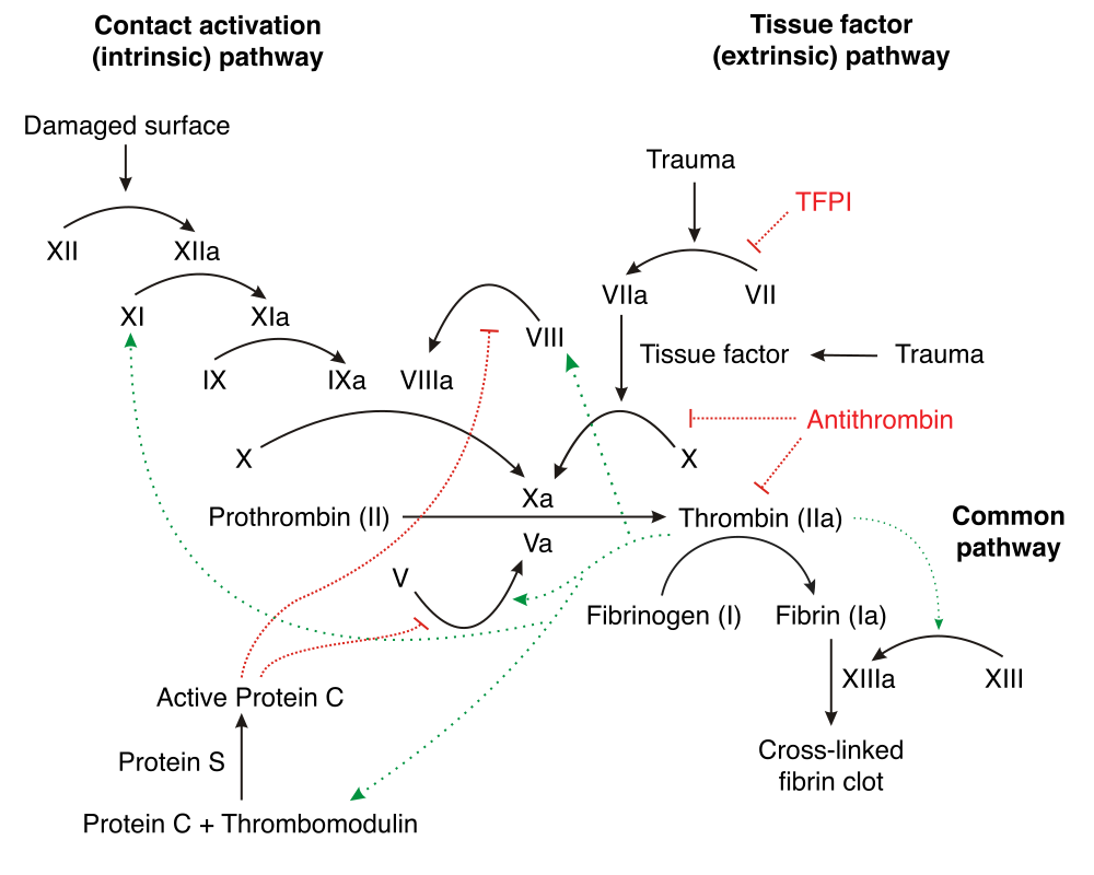
- PT/INR → extrinsic (VII) + common (X, V, II, fibrinogen); monitors warfarin; rises in vitamin K deficiency & liver disease
- aPTT → intrinsic (XII, XI, IX, VIII) + common; monitors unfractionated heparin; prolonged in hemophilia, vWD, lupus anticoagulant
- Factor VII has the shortest half-life (~4-6 h) → PT/INR is the FIRST to rise in early warfarin use, vitamin K deficiency, and early liver disease
- Isolated ↑aPTT that CORRECTS on mixing = factor deficiency; FAILS to correct = inhibitor
- Normal PT AND aPTT but bleeding → think platelet/vWF defect, or factor XIII deficiency (screen: urea clot solubility)
- Factor XII deficiency → ↑aPTT but NO bleeding (asymptomatic lab finding)
- Lupus anticoagulant: ↑aPTT that won't correct, yet causes thrombosis + fetal loss, not bleeding
| PT | aPTT | Think of |
|---|---|---|
| Normal | ↑ | Hemophilia A (VIII)/B (IX), vWD, factor XI or XII def, heparin, lupus anticoagulant |
| ↑ | Normal | Early warfarin, early vitamin K deficiency, factor VII def, early liver disease |
| ↑ | ↑ | DIC, advanced liver disease, late vitamin K def, warfarin overdose, heparin excess, common-pathway (X/V/II/fibrinogen) defect, DOACs |
| Normal | Normal | Platelet dysfunction, mild vWD, factor XIII def, vascular/scurvy |
Vignette A: A 5-year-old boy has recurrent hemarthrosis and deep-muscle bleeds. Labs: normal PT, prolonged aPTT, normal platelets. A 1:1 mixing study corrects the aPTT. Dx / next step: Factor deficiency → order factor VIII and IX activity assays → hemophilia A (VIII) or B (IX), X-linked recessive. Treat acute bleed with factor concentrate; mild hemophilia A can respond to DDAVP (desmopressin), which releases stored factor VIII/vWF.
Vignette B: A 30-year-old woman has two second-trimester miscarriages and a DVT. Labs: prolonged aPTT that does NOT correct on mixing. Dx / next step: Inhibitor → lupus anticoagulant / antiphospholipid syndrome. Confirm with dRVVT plus anticardiolipin and anti-β2-glycoprotein I antibodies (positive on 2 tests ≥12 weeks apart). Despite the long aPTT, the risk is clotting — anticoagulate.
Vignette C: A 62-year-old with gram-negative sepsis oozes from every IV site. Labs: ↑PT, ↑aPTT, ↓platelets, ↓fibrinogen, ↑↑D-dimer, and schistocytes on smear. Dx / next step: DIC. The key move is to treat the underlying trigger (sepsis); support with cryoprecipitate (for fibrinogen <100 mg/dL), FFP, and platelet transfusion only if actively bleeding or high-risk. Board discriminator: DIC vs liver disease — factor VIII is LOW in DIC (consumed) but normal/high in liver disease (made by endothelium, not hepatocytes), helping separate them. Vitamin K deficiency corrects with vitamin K; hepatic synthetic failure does not.

- "1972" = the vitamin K–dependent factors: II, VII, IX, X (plus anticoagulant proteins C and S) — the ones warfarin knocks out
- WEPT: Warfarin → Extrinsic → PT/INR. Its counterpart: heparin → aPTT (intrinsic)
- Extrinsic = one factor (VII), easy to hold in mind; the intrinsic club is the crowd of numbers: XII, XI, IX, VIII
- Protein C has a short half-life and falls fastest when warfarin is started → brief hypercoagulable window → warfarin-induced skin necrosis; overlap (bridge) with heparin until the INR is therapeutic to avoid it
| Drug | Monitor with | Reversal |
|---|---|---|
| Warfarin | PT / INR | Vitamin K; 4-factor PCC for major bleed |
| UF heparin | aPTT (or anti-Xa) | Protamine sulfate |
| LMWH | anti-Xa (aPTT unreliable) | Protamine (partial) |
| Dabigatran (direct IIa) | Dilute thrombin time | Idarucizumab |
| Rivaroxaban/apixaban (Xa) | anti-Xa (drug-calibrated) | Andexanet alfa or PCC |
Elevated INR on warfarin — pick the NEXT BEST STEP:
- INR supratherapeutic but <4.5, no bleeding → lower or hold a dose; recheck
- INR 4.5-10, no bleeding → hold warfarin; routine vitamin K not recommended
- INR >10, no bleeding → hold warfarin + give oral vitamin K
- Serious / life-threatening bleeding (any INR) → 4-factor PCC + IV vitamin K (use FFP only if PCC unavailable)
- In a mechanical-valve patient, avoid over-reversal — weigh thrombosis risk before giving vitamin K
- Isolated ↑PT in a hospitalized patient on broad-spectrum antibiotics + poor intake → vitamin K deficiency; give vitamin K and reassess
Practice Clinical Diagnosis now
Board-style questions, spaced-repetition flashcards, and a Socratic AI tutor — free to start.