CNS Tumors
A boards-focused tour of CNS tumors — from metastases and glioblastoma in adults to posterior-fossa pediatric tumors — pairing classic histology/imaging buzzwords with localization and next-best-step decisions. Emphasizes high-yield distinctions (medulloblastoma vs pilocytic astrocytoma, toxoplasmosis vs primary CNS lymphoma) and management pearls.
Overview & mass effect
Metastases are the most common intracranial tumors in adults (from lung > breast > melanoma > renal > colon) — classically multiple, well-circumscribed lesions at the gray–white junction. Among primary CNS tumors, meningioma is the most common overall (benign) and glioblastoma the most common malignant. Location tracks with age: adult primaries are usually supratentorial; pediatric primaries are usually infratentorial (posterior fossa). Primary CNS tumors almost never metastasize outside the CNS.
Whatever the type, tumors cause disease through mass effect and ↑ intracranial pressure (ICP): headache that is worse in the morning / when lying down, nausea and vomiting, papilledema, and ultimately herniation. Two other flags are new-onset seizures and focal deficits that localize the lesion (e.g., cerebellar mass → ataxia; chiasm → bitemporal hemianopia). Vasogenic edema surrounds the mass. Contrast-enhanced MRI is the imaging study of choice.
- Glioblastoma (astrocytoma, WHO grade IV): most common 1° malignant brain tumor; "butterfly" spread across the corpus callosum; ring-enhancing with central necrosis; pseudopalisading necrosis; GFAP+; IDH-wildtype; dismal prognosis (~1 yr).
- Meningioma: 2nd most common; women; from arachnoid cap cells; extra-axial with a dural tail; psammoma bodies + whorls; often silent or causes seizures; benign.
- Vestibular schwannoma: cerebellopontine angle, CN VIII; sensorineural hearing loss + tinnitus; S-100+; bilateral = NF2.
- Oligodendroglioma: frontal lobe; "fried-egg" cells + "chicken-wire" capillaries; often calcified; 1p/19q co-deletion → chemo/radiosensitive, better prognosis.
- Hemangioblastoma: cerebellar, hypervascular, foamy stromal cells; VHL; secretes EPO → secondary polycythemia.
- Pituitary adenoma (prolactinoma most common): bitemporal hemianopia.
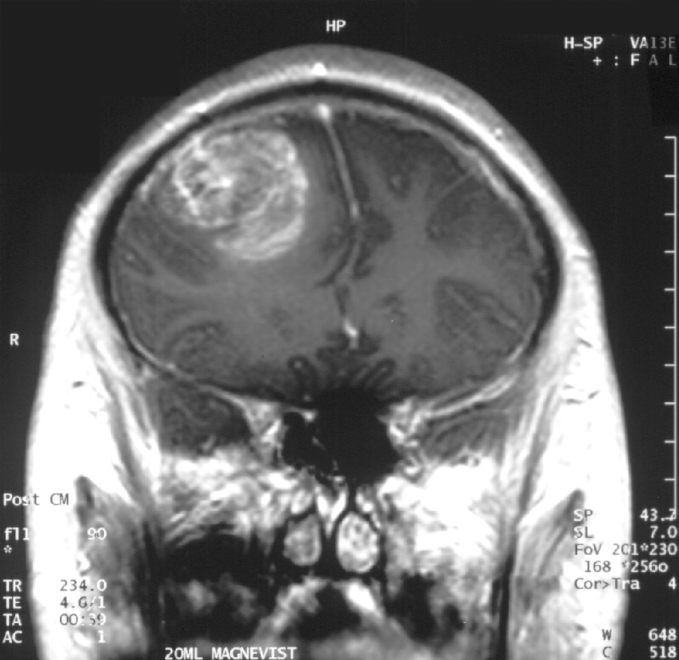
- Pilocytic astrocytoma: most common pediatric CNS tumor; cerebellum; cystic mass with an enhancing mural nodule; Rosenthal fibers + eosinophilic granular bodies; GFAP+; benign, excellent prognosis; NF1-associated (optic nerve glioma).
- Medulloblastoma: most common malignant peds tumor; cerebellar vermis → truncal ataxia + obstructive hydrocephalus; small round blue cells, Homer-Wright rosettes; seeds CSF → "drop metastases"; radiosensitive.
- Ependymoma: 4th ventricle → hydrocephalus; perivascular pseudorosettes; rod-shaped blepharoplasts.
- Craniopharyngioma: suprasellar; remnant of Rathke pouch; calcified; "motor-oil" cyst fluid with cholesterol crystals; bitemporal hemianopia; most common childhood supratentorial tumor.
Rule of thumb: kids' tumors cluster in the posterior fossa → cerebellar signs + hydrocephalus.
Comparison of key CNS tumors
| Tumor | Location | Histology / imaging buzzword | Pearl |
|---|---|---|---|
| Glioblastoma | Hemispheres; crosses corpus callosum | Ring-enhancing + pseudopalisading necrosis; GFAP+ | Most common 1° malignant |
| Meningioma | Extra-axial, dura-based | Dural tail; psammoma bodies; whorls | Women; benign; seizures |
| Vestibular schwannoma | CP angle (CN VIII) | S-100+; Antoni A & B | SNHL + tinnitus; bilateral = NF2 |
| Oligodendroglioma | Frontal lobe | Fried-egg cells; chicken-wire vessels; calcified | 1p/19q co-del → chemosensitive |
| Pilocytic astrocytoma | Cerebellum (child) | Cystic + mural nodule; Rosenthal fibers | Most common peds tumor; benign |
| Medulloblastoma | Cerebellar vermis (child) | Small blue cells; Homer-Wright rosettes | Drop mets; radiosensitive |
| Ependymoma | 4th ventricle (child) | Perivascular pseudorosettes; blepharoplasts | Hydrocephalus |
| Craniopharyngioma | Suprasellar | Calcified; "motor-oil" cyst | Rathke pouch; bitemporal hemianopia |
| Hemangioblastoma | Cerebellum | Foamy cells; hypervascular | VHL; EPO → polycythemia |
Vignette: A 6-year-old has 3 weeks of morning headaches and vomiting and is unsteady with a wide-based gait and truncal titubation (sways when sitting/standing). Exam shows papilledema and truncal ataxia. MRI reveals a midline cerebellar (vermian) mass filling the 4th ventricle with hydrocephalus.
Diagnosis: Medulloblastoma — a solid, contrast-enhancing midline mass. Contrast this with a well-circumscribed, off-midline (hemispheric) cystic mass with an enhancing mural nodule, which points to pilocytic astrocytoma.
Next best step: MRI of the entire neuraxis (brain + spine) to detect CSF "drop metastases," followed by maximal safe resection (then craniospinal radiation/chemo). Do NOT perform LP first — a posterior fossa mass with ↑ICP risks cerebellar tonsillar herniation.

Vignette: A man with AIDS (CD4 40), off prophylaxis, has headache and focal weakness. MRI shows multiple ring-enhancing lesions in the basal ganglia and at the gray–white junction.
Two big differentials: Toxoplasmosis (usually multiple, ring-enhancing) vs primary CNS lymphoma (EBV-driven, often single/periventricular, may ring-enhance from central necrosis in AIDS). Distinguishers favoring lymphoma: positive CSF EBV DNA and increased uptake on thallium-201 SPECT / PET.
Next best step: start empiric pyrimethamine + sulfadiazine for toxoplasmosis and repeat imaging in ~2 weeks. Lesions shrink → toxoplasmosis. No response → stereotactic brain biopsy (primary CNS lymphoma → high-dose methotrexate). Avoid empiric steroids before biopsy if lymphoma is suspected — they transiently shrink it and can obscure the diagnosis.
Laminated, concentric calcifications (psammoma bodies) — a meningioma hallmark — also appear in a classic quartet. Remember PSaMMoma:
- P — Papillary carcinoma of the thyroid
- S — Serous papillary cystadenocarcinoma of the ovary (and serous endometrial carcinoma)
- M — Meningioma
- M — Mesothelioma

- Imaging: contrast-enhanced MRI is first-line; definitive diagnosis needs tissue (biopsy/resection).
- ↑ICP / posterior fossa mass: image first — do not LP (herniation risk). Treat vasogenic edema with dexamethasone; control seizures with antiepileptics.
- Glioblastoma: maximal safe resection + radiation + temozolomide (Stupp protocol); MGMT promoter methylation predicts temozolomide benefit.
- Prolactinoma: dopamine agonist (cabergoline/bromocriptine) first-line, not surgery.
- Metastasis: most common intracranial tumor overall — search for the primary (lung, breast, melanoma, renal, colon).
- Phakomatoses: NF1 → optic glioma / pilocytic astrocytoma; NF2 → bilateral vestibular schwannomas, meningiomas, ependymomas; VHL → hemangioblastoma (+ RCC, pheo); tuberous sclerosis → subependymal giant cell astrocytoma (SEGA).
Practice Neuro now
Board-style questions, spaced-repetition flashcards, and a Socratic AI tutor — free to start.