Cerebral Vascular Territories & the Circle of Willis
A high-yield Step 1 neuroanatomy lesson mapping the Circle of Willis and the ACA/MCA/PCA plus vertebrobasilar territories directly onto the deficits the boards test — cortical stroke patterns, brainstem syndromes, and the aneurysm/hemorrhage correlations. Built around the rule that knowing which artery feeds which cortex lets you predict every lesion.
The Two Circulations and the Ring That Joins Them
The brain is perfused by two paired arterial systems that meet in an anastomotic ring at the base of the brain — the Circle of Willis.
- Anterior circulation arises from the internal carotid arteries (ICA), which give off the anterior cerebral (ACA) and middle cerebral (MCA) arteries.
- Posterior circulation arises from the paired vertebral arteries, which fuse into the basilar artery and terminate as the posterior cerebral arteries (PCA).
The ring is closed by three communicating vessels: a single anterior communicating artery (ACOM) bridging the two ACAs, and paired posterior communicating arteries (PCOM) linking each ICA to its PCA. This collateral loop can cross-fill a stenosed vessel — but its junctions are also the classic sites of saccular (berry) aneurysms. On the boards, almost every question reduces to one idea: know which artery feeds which piece of cortex, and you can predict the deficit.
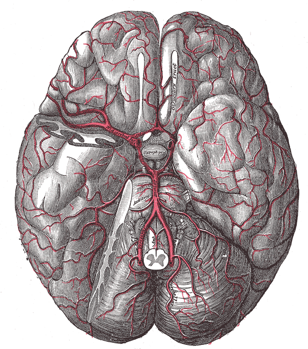
- Anterior circulation = ICA → ACA + MCA; posterior = vertebral → basilar → PCA
- ACOM joins the two ACAs; paired PCOM join each ICA to its PCA — together they complete the ring
- ACA = contralateral LEG (medial motor/sensory strip, paracentral lobule) ± abulia, urinary incontinence
- MCA = contralateral FACE + ARM + language (dominant/left) or neglect (nondominant/right); the most commonly occluded cerebral artery
- PCA = occipital lobe → contralateral homonymous hemianopia with MACULAR SPARING
- Lenticulostriate arteries (deep branches off proximal MCA) feed the internal capsule / basal ganglia → lacunar strokes (chronic HTN)
- ACOM = most common berry-aneurysm site; PCOM aneurysm → CN III palsy ("down-and-out" eye, blown pupil)
- Watershed zones (ACA–MCA, MCA–PCA borders) infarct in severe hypotension → bilateral proximal ("man-in-a-barrel") weakness
Cerebral Artery Territories → Deficits
| Artery | Cortex / structures supplied | Classic deficit if occluded |
|---|---|---|
| ACA | Medial frontal & parietal lobes (leg–foot homunculus, paracentral lobule) | Contralateral lower-limb motor/sensory loss; abulia, urinary incontinence |
| MCA | Lateral hemisphere (face–arm homunculus; Broca & Wernicke) | Contralateral face + arm motor/sensory loss; aphasia (dominant/left) or hemineglect (nondominant/right) |
| Lenticulostriate (deep branches of proximal MCA / M1) | Internal capsule, basal ganglia (putamen, globus pallidus, caudate) | Lacunar pure-motor hemiparesis (posterior limb of internal capsule); classic site of hypertensive (Charcot-Bouchard) hemorrhage |
| PCA | Occipital cortex, inferior temporal lobe, thalamus (incl. VPL via thalamoperforators), midbrain | Contralateral homonymous hemianopia with macular sparing; thalamoperforator lacune → pure-sensory stroke |

Vignette 1 — ACA: A 70-year-old has sudden weakness and numbness of the right leg, with arm and face spared; he is apathetic and incontinent. → Left ACA infarct (medial homunculus = lower limb).
Vignette 2 — MCA (dominant): A hypertensive man has right face and arm weakness and cannot produce fluent speech, though he follows commands. → Left MCA infarct involving Broca area (nonfluent aphasia, comprehension intact); face/arm > leg because the MCA serves the lateral homunculus.
Vignette 3 — MCA (nondominant): A woman denies her left arm is weak and ignores the left half of her plate. → Right (nondominant) MCA infarct → hemispatial neglect.
Vignette 4 — PCA: After a cardioembolic event, a patient has lost the right half of the visual field in both eyes but still reads the central letters. → Left PCA (occipital) infarct → right homonymous hemianopia with macular sparing (occipital pole receives collateral MCA supply).

- Homunculus rule: ACA = leg, MCA = face + arm. The medial strip (ACA) is the foot/leg dangling into the interhemispheric fissure; the big lateral "middle" of the body map (MCA) is face and arm.
- "Facial droop means AICA's pooped" — AICA lateral pontine stroke → ipsilateral facial paralysis (CN VII) + deafness (labyrinthine artery / CN VIII).
- "Don't PICA horse that can't eat" — PICA lateral medullary (Wallenberg) → dysphagia / hoarseness (nucleus ambiguus, CN IX/X).
- PCOM → CN III: a Posterior communicating aneurysm compresses the adjacent third nerve → "down-and-out" eye, ptosis, blown (dilated) pupil — because the parasympathetic fibers ride the outside of CN III and are squeezed first.
PCOM aneurysm → CN III palsy: A woman with a thunderclap headache has a fixed, dilated pupil and a "down-and-out" eye with ptosis. Compression hits the superficial parasympathetic fibers first (pupil blown) — the mirror image of an ischemic/diabetic CN III palsy, which is pupil-SPARING.
ACOM aneurysm → subarachnoid hemorrhage: The anterior communicating artery is the single most common berry-aneurysm site. Rupture → SAH: "worst headache of my life," nuchal rigidity, xanthochromia on LP. A large unruptured ACOM aneurysm can compress the nearby optic chiasm and cause visual-field defects. Berry aneurysms associate with ADPKD and vascular (type IV) Ehlers-Danlos, plus modifiable risk factors — smoking, hypertension, advancing age.
Charcot-Bouchard microaneurysms (a DIFFERENT lesion) form on lenticulostriate vessels in chronic hypertension and rupture into the basal ganglia (putamen) — an intraparenchymal, not subarachnoid, bleed.
Vertebrobasilar / Brainstem Stroke Syndromes
| Syndrome (artery) | Structures hit | Key deficits |
|---|---|---|
| Medial medullary (anterior spinal a. / vertebral branches) | Pyramid (corticospinal), medial lemniscus, CN XII | Contralateral arm/leg weakness + loss of vibration/proprioception; ipsilateral tongue deviation |
| Lateral medullary / Wallenberg (PICA) | Nucleus ambiguus, spinothalamic, spinal trigeminal, sympathetics, vestibular, inferior cerebellum | Dysphagia/hoarseness; ipsilateral face & contralateral body pain/temp loss; Horner; vertigo, ipsilateral ataxia |
| Lateral pontine (AICA) | CN VII & VIII nuclei, spinothalamic, spinal trigeminal, sympathetics, cerebellum | Ipsilateral facial paralysis + deafness; ipsilateral face & contralateral body pain/temp loss; Horner, ataxia |
- Watershed (border-zone) infarcts occur between the ACA–MCA and MCA–PCA territories during global hypoperfusion (sepsis, cardiac arrest, hemorrhagic shock)
- Clue: bilateral proximal-limb (shoulder/hip) weakness — the "man in a barrel"; posterior watershed adds visual deficits
- Lacunar infarcts = occlusion of small lenticulostriate/thalamoperforator vessels from chronic HTN + diabetes (lipohyalinosis):
- Posterior limb of internal capsule → pure motor hemiparesis
- VPL of thalamus (thalamoperforators) → pure sensory stroke
- Basis pontis / internal capsule → dysarthria–clumsy hand or ataxic hemiparesis
- The lateral lenticulostriate arteries are Charcot's "arteries of cerebral hemorrhage" — small, penetrating end-arteries unforgiving of chronic hypertension
Practice Anatomy now
Board-style questions, spaced-repetition flashcards, and a Socratic AI tutor — free to start.