Cardiomyopathies: Dilated, Hypertrophic & Restrictive
A board-focused walkthrough of the three cardiomyopathies—dilated, hypertrophic, and restrictive—linking pathophysiology to the classic vignette buzzwords, ECG/echo findings, and next-best-step decisions. Includes HCM murmur maneuvers, the cardiac-amyloid voltage–mass clue, and the restrictive-vs-constrictive discriminator.
Overview: three phenotypes, one board skill
Cardiomyopathies are intrinsic diseases of heart muscle causing mechanical/electrical dysfunction that is not explained by coronary, valvular, hypertensive, or congenital disease. Boards sort them into three functional phenotypes:
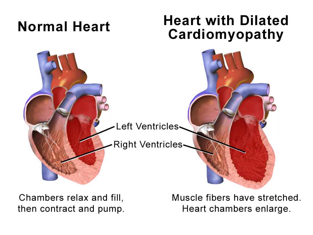
- Dilated (DCM) — a systolic (pump) problem: the ventricle dilates and thins, so contractility and ejection fraction fall. Most common cardiomyopathy and the leading indication for heart transplant.
- Hypertrophic (HCM) — primarily a diastolic (filling) problem: a thick, stiff, often asymmetrically hypertrophied septum; autosomal-dominant sarcomere mutations (β-myosin heavy chain MYH7, myosin-binding protein C MYBPC3). The classic board answer for sudden cardiac death in young athletes.
- Restrictive (RCM) — also diastolic, but from a rigid, non-compliant myocardium; wall thickness and EF are near-normal early. Least common; usually infiltrative (amyloid, sarcoid, hemochromatosis).
The unifying skill the exam tests: match the vignette's buzzwords to a phenotype, then choose the next best step.
- DCM = eccentric hypertrophy (sarcomeres added in series): ↓EF, dilated chambers, S3 gallop, functional MR, mural thrombus → systemic emboli.
- HCM = concentric/asymmetric hypertrophy (sarcomeres in parallel): preserved/↑EF, S4, dynamic LVOT obstruction from SAM (systolic anterior motion of the mitral valve) — present in only a subset (obstructive HCM).
- HCM murmur: harsh crescendo–decrescendo at the left lower sternal border; louder with ↓preload (Valsalva, standing) and softer with ↑preload/afterload (squatting, handgrip) — the opposite of aortic stenosis.
- RCM: near-normal EF and wall size, biatrial enlargement, high filling pressures; triggers = amyloid, sarcoid, hemochromatosis, endomyocardial fibrosis, Löffler.
- Echocardiography is the initial test of choice for all three.
- Cardiac amyloid clue: low-voltage ECG with thick walls on echo (voltage–mass mismatch) plus a pseudoinfarct (Q-wave) pattern.
Side-by-side comparison
| Feature | Dilated | Hypertrophic | Restrictive |
|---|---|---|---|
| Core defect | Systolic (↓contractility) | Diastolic (impaired filling ± dynamic LVOT obstruction) | Diastolic (stiff wall) |
| LV cavity / walls | Dilated, thin | Small cavity, thick septum | Normal size, near-normal wall* |
| EF | ↓ | Preserved/↑ | Preserved early |
| Extra heart sound | S3 | S4 | S3/S4 |
| Classic causes | Idiopathic/genetic (titin), alcohol, myocarditis (Coxsackie B), peripartum, doxorubicin, cocaine, Chagas, wet beriberi | AD sarcomere mutation (MYH7, MYBPC3) | Amyloid, sarcoid, hemochromatosis, endomyocardial fibrosis, Löffler, radiation |
| Management | HFrEF GDMT (ARNI/ACEi, β-blocker, MRA, SGLT2i); ICD if EF ≤35% despite ≥3 mo GDMT | β-blocker/verapamil; avoid preload/afterload reduction; ICD if high SCD risk; myectomy/ablation | Treat the cause; cautious diuresis; avoid digoxin in amyloid |
Vignette: An 18-year-old basketball player collapses during a game. He reports prior exertional syncope and a family history of sudden death. Exam: a harsh systolic murmur at the LLSB that intensifies with Valsalva and softens with squatting.
Diagnosis: Hypertrophic cardiomyopathy with dynamic LVOT obstruction.
Next steps / management:
- Transthoracic echo — asymmetric septal hypertrophy, SAM, measurable LVOT gradient.
- ECG — LVH, deep narrow septal Q waves ± giant T-wave inversions (apical variant).
- Restrict intense competitive athletics; keep well hydrated (avoid hypovolemia).
- β-blocker (or verapamil) first-line; disopyramide or mavacamten (cardiac myosin inhibitor) for refractory obstruction.
- ICD for high SCD risk (prior arrest/sustained VT, family Hx SCD, unexplained syncope, septum ≥30 mm, apical aneurysm, extensive LGE).
- Avoid nitrates, dihydropyridine CCBs, ACE inhibitors, aggressive diuresis, and digoxin — ↓preload/afterload or ↑contractility worsens obstruction.
- Screen first-degree relatives.

Vignette: A 72-year-old man has progressive HFpEF, bilateral carpal tunnel syndrome, and orthostatic hypotension. ECG shows low voltage, yet echo shows thickened ventricular walls with a granular/speckled myocardium and biatrial enlargement.
Diagnosis: Restrictive cardiomyopathy from cardiac amyloidosis — the voltage–mass discordance (low voltage + thick walls) is the giveaway.
Next steps:
- Serum/urine free light chains + SPEP/UPEP to screen for AL amyloid.
- If light chains are negative → technetium-99m pyrophosphate (PYP) scan for ATTR amyloid.
- Endomyocardial biopsy if needed: apple-green birefringence under polarized light with Congo red stain.
- Treat by type: AL → chemotherapy; ATTR → tafamidis (transthyretin stabilizer).
- Avoid digoxin — it binds amyloid fibrils, causing enhanced sensitivity and toxicity. Non-dihydropyridine CCBs and β-blockers are poorly tolerated in a fixed-output, infiltrated ventricle (hypotension, bradycardia) — use with caution.
HCM = "SAM" — Systolic Anterior Motion of the mitral valve: the mechanism of dynamic LVOT obstruction (and of the accompanying mitral regurgitation).
Maneuver rule (highest-yield): HCM and mitral valve prolapse are the classic pair whose murmurs get louder with ↓preload (Valsalva, standing) — nearly every other left-sided murmur gets softer. Squatting/handgrip (↑preload/afterload) do the reverse.
New DCM → hunt the reversible cause (finding and treating it can recover EF): alcohol, thiamine deficiency (wet beriberi), tachycardia-mediated, thyroid disease, cocaine, peripartum, viral myocarditis (Coxsackie B), and anthracyclines (doxorubicin).
Restrictive cardiomyopathy vs constrictive pericarditis
This is a favorite Step 2 CK discriminator because both cause right-sided diastolic failure with elevated, equalized filling pressures, Kussmaul sign, and prominent x/y descents.
Points to constrictive pericarditis (the surgically curable one):
- History of TB, prior cardiac surgery, or chest radiation
- Pericardial calcification/thickening on CXR or CT
- Pericardial knock and respirophasic ventricular interdependence (septal bounce; discordant LV/RV filling with respiration)
- Relatively normal/low BNP; structurally normal myocardium
Points to restrictive cardiomyopathy:
- Markedly elevated BNP, thick/infiltrated myocardium, biatrial enlargement
- No pericardial calcification; ventricular filling is concordant with respiration
Management diverges sharply: pericardiectomy can cure constriction, whereas RCM is managed by treating the underlying infiltrative disease.
- Peripartum cardiomyopathy: a DCM appearing from the last month of pregnancy to ~5 months postpartum; treat as HFrEF but hold ACEi/ARB/ARNI/SGLT2i/MRA while pregnant or breastfeeding — use hydralazine + nitrate and a β-blocker; many patients recover EF.
- Takotsubo (stress) cardiomyopathy: after intense emotional/physical stress in older (typically post-menopausal) women; apical ballooning with normal coronaries; mimics STEMI but is usually reversible with supportive care.
- DCM GDMT (four pillars): ARNI (or ACEi/ARB) + evidence-based β-blocker + MRA + SGLT2 inhibitor; ICD if EF ≤35% despite ≥3 months of GDMT; anticoagulate for LV thrombus or atrial fibrillation.
- Sarcoid RCM: patchy infiltration → high-grade AV block/VT; treat with corticosteroids ± device.
- Hemochromatosis can produce both restrictive and dilated patterns; treat with phlebotomy/chelation.
Practice Cardiology now
Board-style questions, spaced-repetition flashcards, and a Socratic AI tutor — free to start.