Cardiac Embryology & Congenital Heart Defects
Traces cardiac development from the splanchnic-mesoderm heart tube through looping and septation, mapping each embryonic segment to its adult derivative, then ties the classic Step 1 congenital defects to the exact septation and neural-crest errors that cause them.
The developmental sequence
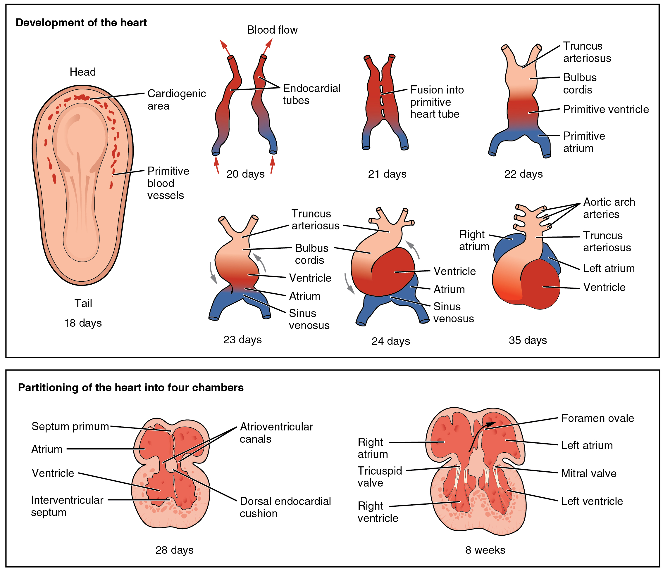
The heart is the first functional organ, beating around day 22 (start of week 4). It arises from splanchnic (lateral plate) mesoderm in the cardiogenic field. Paired endocardial (heart) tubes fuse into a single primitive heart tube, which develops five dilations you must know cranial → caudal: truncus arteriosus → bulbus cordis → primitive ventricle → primitive atrium → sinus venosus.
During cardiac looping (days 23–28), normal dextral (D-)looping swings the bulbus cordis ventrally and to the right while the primitive ventricle shifts left, establishing correct left–right chamber topology. Abnormal L-looping → dextrocardia. Because looping and septation occur in weeks 3–8, this is the critical window in which teratogens and most structural defects arise.
- First functional organ; beats ~day 22; derived from splanchnic (lateral plate) mesoderm.
- Heart-tube segments cranial→caudal: Truncus arteriosus → Bulbus cordis → primitive Ventricle → primitive Atrium → Sinus venosus.
- Cardiac neural crest cells populate the truncal/bulbar ridges to septate the outflow tract → failure causes conotruncal defects (persistent truncus, TGA, TOF); linked to 22q11.2 deletion (DiGeorge).
- Endocardial cushions form the AV valves, the membranous interventricular septum, and part of the atrial septum → defects cause ostium primum ASD / AV septal defect (Down syndrome).
- Septation (atrial, ventricular, outflow) completes by the end of week 7–8; abnormal looping → dextrocardia.
- The semilunar (aortic/pulmonary) valves derive from the outflow-tract (conotruncal) cushions, not the AV cushions.
- VSD (usually membranous) is the most common congenital heart defect overall.
Heart-tube segment → adult derivative
| Embryonic structure | Adult derivative |
|---|---|
| Truncus arteriosus | Ascending aorta + pulmonary trunk |
| Bulbus cordis | Smooth outflow (conus) parts of ventricles — RV infundibulum/conus arteriosus, LV aortic vestibule |
| Primitive ventricle | Trabeculated (rough) parts of left & right ventricles |
| Primitive atrium | Trabeculated parts of left & right atria (auricles / pectinate muscle) |
| Right horn of sinus venosus | Smooth part of right atrium (sinus venarum) |
| Left horn of sinus venosus | Coronary sinus |
| Right common + anterior cardinal veins | SVC |
| Incorporated pulmonary veins | Smooth part of left atrium |
Three septation events
1. Atrial septum. Septum primum grows down toward the endocardial cushions (leaving the ostium primum). Before it seals, apoptosis opens the ostium secundum in its upper part. Septum secundum then grows down to the right of septum primum, leaving the foramen ovale, with the remaining septum primum acting as its one-way flap valve. At birth, rising LA pressure presses the flap shut → the fossa ovalis. Failure to fuse = patent foramen ovale.
2. Ventricular septum. A muscular portion grows upward from the apex; the membranous portion is completed by the endocardial cushions and conotruncal ridges — and closes last.
3. Outflow tract (conotruncal). Cardiac neural crest cells fill the truncal and bulbar ridges, which spiral and fuse into the aorticopulmonary (spiral) septum, dividing the truncus into aorta and pulmonary trunk. The spiral course explains why the great vessels twist around each other.
Defect → embryologic mechanism
| Congenital defect | Mechanism |
|---|---|
| Persistent truncus arteriosus | Aorticopulmonary (neural-crest) septum fails to form → single trunk; almost always with a VSD |
| D-Transposition of great arteries | Aorticopulmonary septum fails to spiral (grows straight) → aorta off RV, PA off LV |
| Tetralogy of Fallot | Anterosuperior deviation of the infundibular (outlet) septum → RV outflow obstruction |
| Ostium secundum ASD | Excess resorption of septum primum or deficient septum secundum (most common ASD) |
| Ostium primum ASD / AVSD | Endocardial cushions fail to fuse (Down syndrome) |
| Membranous VSD | Membranous IV septum fails to close (most common VSD; VSD = most common CHD overall) |
| Patent ductus arteriosus | Left 6th-arch–derived ductus fails to close; assoc. congenital rubella |
| Patent foramen ovale | Septum primum & secundum fail to fuse after birth |
A 4-month-old boy has recurrent spells of turning blue during crying and feeds that resolve when his mother pulls his knees to his chest. Exam: harsh systolic ejection murmur at the left upper sternal border. CXR shows a boot-shaped heart.
Diagnosis: Tetralogy of Fallot — the most common cyanotic CHD presenting beyond the neonatal period.
- Single cause: anterosuperior deviation of the infundibular septum, producing all four features — pulmonary/RV outflow stenosis, overriding aorta, VSD, and RVH (boot-shaped heart).
- Degree of pulmonary stenosis determines severity and the size of the right-to-left shunt.
- "Tet spells": squatting or knee-chest position ↑ systemic vascular resistance → ↓ right-to-left shunt → improves cyanosis.

A term newborn of a mother with poorly controlled diabetes becomes deeply cyanotic within hours of birth, and the cyanosis does not improve with supplemental O₂. CXR: narrow mediastinum — "egg on a string."
Diagnosis: D-Transposition of the great arteries — the most common cyanotic CHD presenting at birth.
- Mechanism: the aorticopulmonary septum fails to spiral, so the aorta arises from the RV and the pulmonary artery from the LV → two parallel circuits.
- Incompatible with life unless the circuits mix through a PFO/ASD, VSD, or PDA.
- Management: prostaglandin E1 to keep the ductus arteriosus open (± balloon atrial septostomy) as a bridge to the arterial switch operation.
5 T's of cyanotic congenital heart disease — numbered by the trick that names them:
- Truncus arteriosus — 1 vessel
- Transposition of the great arteries — 2 vessels switched
- Tricuspid atresia — 3 (tri = 3)
- Tetralogy of Fallot — 4 features
- TAPVR (total anomalous pulmonary venous return) — 5 letters
PROVe — the four features of Tetralogy of Fallot:
- Pulmonary stenosis (RV outflow obstruction — most important)
- Right ventricular hypertrophy (boot-shaped heart)
- Overriding aorta
- VSD
- Three fetal shunts: ductus venosus (umbilical vein → IVC, bypasses liver), foramen ovale (RA → LA), ductus arteriosus (pulmonary trunk → aorta, bypasses lungs).
- Ductus arteriosus derives from the left 6th aortic arch; kept open by prostaglandins (PGE1/E2), closed by indomethacin → becomes the ligamentum arteriosum.
- Aortic arch derivatives: 3rd → common carotid + proximal internal carotid; 4th → aortic arch (left) & proximal right subclavian (right); 6th → proximal pulmonary arteries + ductus arteriosus (left).
- Coarctation of the aorta: narrowing near the ligamentum arteriosum; assoc. Turner syndrome and bicuspid aortic valve.
- Fetal remnants: foramen ovale → fossa ovalis; ductus venosus → ligamentum venosum; umbilical vein → ligamentum teres hepatis (round ligament); ductus arteriosus → ligamentum arteriosum.
Practice Embryology now
Board-style questions, spaced-repetition flashcards, and a Socratic AI tutor — free to start.