Cardiac Anatomy & Coronary Circulation
A Step 1 high-yield tour of cardiac anatomy — chamber/surface relations, coronary artery territories with their MI/ECG correlations, nodal blood supply and venous drainage, and pericardial anatomy — anchored to the injury and vignette patterns the boards actually test.
Orientation: chambers, surfaces & borders
The heart sits in the middle mediastinum, rotated so that the right ventricle (RV) — not the left — forms most of the anterior (sternocostal) surface, lying directly behind the sternum. Knowing which chamber makes which border explains the classic exam findings:
- Right border = right atrium (RA)
- Left border & apex = left ventricle (LV); the apex sits at the 5th intercostal space, midclavicular line
- Inferior (diaphragmatic) surface = mainly LV, with part of the RV
- Base (posterior surface) = mainly left atrium (LA) — the most posterior chamber
Because the LA is most posterior, it abuts the esophagus and left recurrent laryngeal nerve. LA enlargement (e.g., mitral stenosis) can therefore cause dysphagia and hoarseness (Ortner / cardiovocal syndrome). Because the RV is most anterior, it is the chamber injured first in penetrating sternal trauma.

- Aortic sinuses (of Valsalva) give rise to the coronaries: right cusp → RCA, left cusp → left main, posterior cusp = non-coronary (no artery)
- Coronaries fill during DIASTOLE — systolic myocardial compression squeezes the vessels; tachycardia shortens diastole → subendocardial ischemia (subendocardium is the most vulnerable layer)
- SA node: junction of SVC and RA (upper crista terminalis) — the pacemaker
- AV node: floor of RA at the apex of the triangle of Koch (tendon of Todaro + septal leaflet of tricuspid + os of the coronary sinus)
- Fossa ovalis: interatrial septum; remnant of the foramen ovale — a patent foramen ovale permits paradoxical embolism
- Ligamentum arteriosum (remnant of the ductus arteriosus) tethers the aortic arch to the pulmonary trunk; the left recurrent laryngeal nerve hooks under the arch here
- Posteromedial papillary muscle has a single blood supply (PDA) → prone to rupture in inferior MI
Coronary arteries → territory → occlusion syndrome
| Artery | Origin / course | Supplies | Occlusion → territory (ECG leads) |
|---|---|---|---|
| LAD (anterior interventricular) | From left main, anterior IV groove | Anterior 2/3 of IV septum, anterior LV wall, apex | Anteroseptal MI — V1–V4; the "widowmaker"; most commonly occluded |
| LCX (circumflex) | From left main, left AV groove | Lateral & posterior LV wall; SA node in ~40% | Lateral MI — I, aVL, V5–V6 |
| RCA | Right aortic sinus, right AV groove | RV free wall; SA node (~60%); AV node (if right-dominant) | Inferior MI — II, III, aVF; bradyarrhythmia / AV block; RV infarct |
| PDA (posterior descending) | From RCA in right dominance (~85%) | Inferior LV wall, posterior 1/3 of septum, posteromedial papillary muscle | Parent (dominant) artery defines dominance; the AV-nodal branch arises at the crux from the dominant vessel |
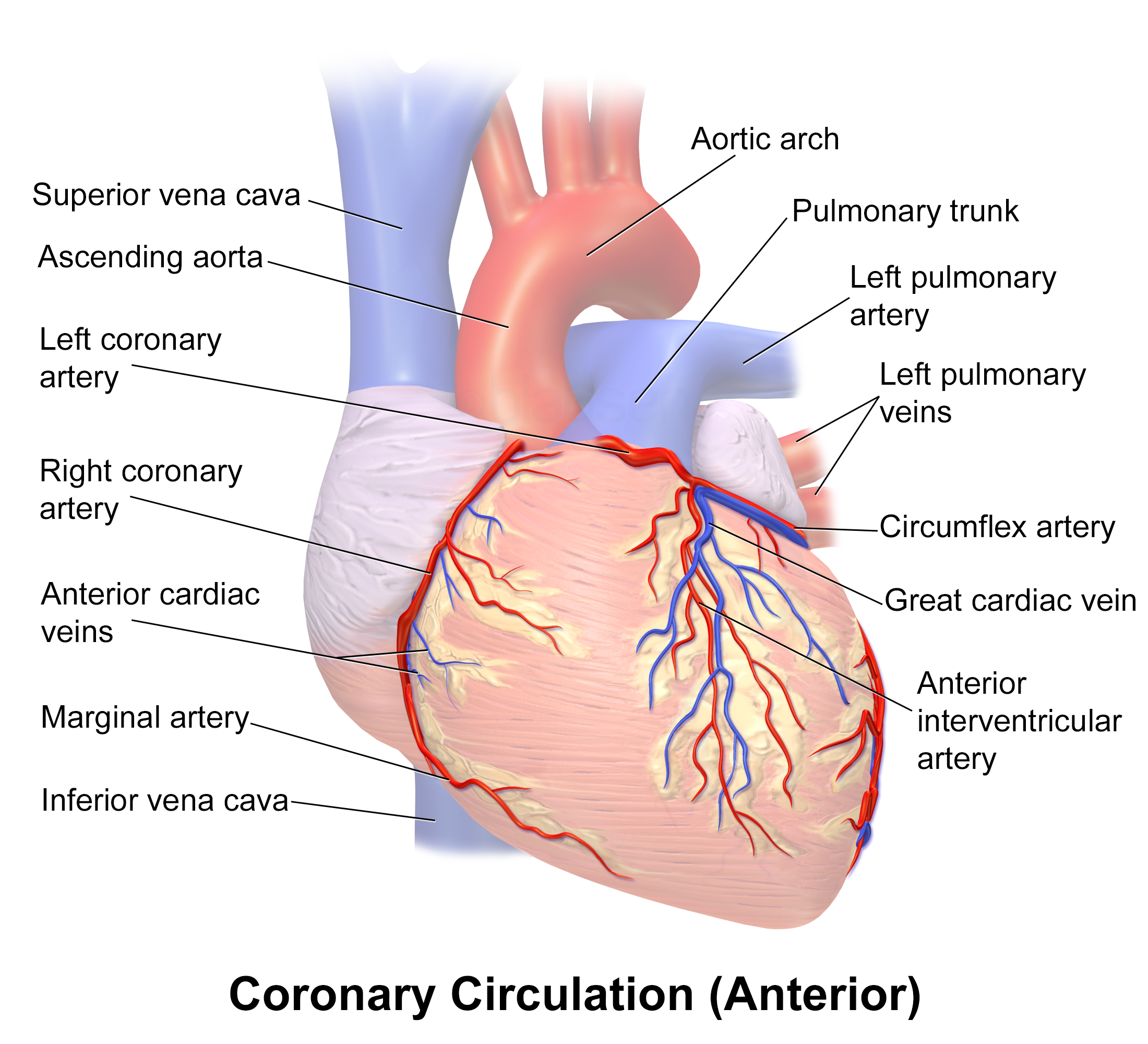
- RCA feeds the nodes → an inferior (RCA) MI classically causes bradycardia and AV block; nitrates are risky if there is RV involvement (RV infarct is preload-dependent → hypotension)
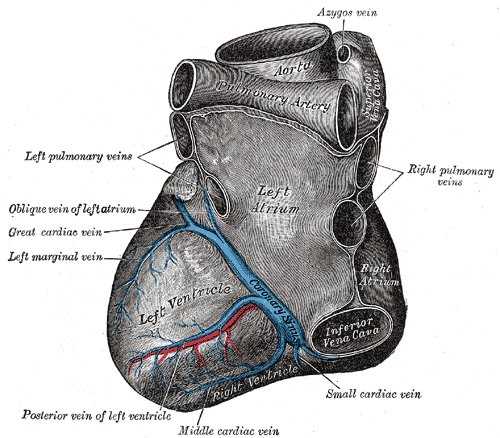
- Coronary sinus runs in the posterior AV groove and empties into the RA (between the IVC orifice and the tricuspid opening, adjacent to the AV node)
- Tributaries: great cardiac vein (with LAD then LCX), middle cardiac vein (with PDA), small cardiac vein
- Anterior cardiac veins drain the RV directly into the RA (bypass the coronary sinus)
- Thebesian (smallest cardiac) veins drain directly into the chambers — a small physiologic venous-admixture shunt
- A dilated coronary sinus is a clue to a persistent left SVC or elevated right-heart pressures
Vignette 1 — 58-y-old man, crushing substernal pain, ST elevation in V1–V4. → LAD occlusion (anteroseptal, the widowmaker), the most commonly occluded coronary.
Vignette 2 — Chest pain with ST elevation in II, III, aVF, plus bradycardia and hypotension. → RCA occlusion (inferior wall). The RCA supplies the AV node (block/bradyarrhythmia) and the RV — give fluids, avoid nitrates.
Vignette 3 — 3–5 days after an inferior MI, a patient develops a new holosystolic murmur and flash pulmonary edema. → Rupture of the posteromedial papillary muscle → acute mitral regurgitation. It ruptures because its single blood supply is the PDA, whereas the anterolateral papillary muscle has dual supply (LAD + LCX).
Vignette 4 — Lateral MI with changes in I, aVL, V5–V6. → LCX occlusion.
Cardiac tamponade — pericardial fluid restricts filling. Look for Beck's triad: hypotension + distended neck veins (JVD) + muffled heart sounds, with pulsus paradoxus (>10 mmHg inspiratory SBP fall) and electrical alternans on ECG. Treat with pericardiocentesis.
Referred pain — two different pathways:
- Pericardium (fibrous + parietal serous) is innervated by the phrenic nerve (C3–C5) → pericarditis pain refers to the shoulder / supraclavicular region; classically worse supine, relieved by leaning forward.
- Cardiac ischemic (visceral afferent) pain travels with sympathetics to T1–T4 → refers to the chest, medial left arm, and jaw.
Surgical landmarks:
- Transverse pericardial sinus lies behind the aorta + pulmonary trunk; a surgeon passes a finger through it to clamp the great arterial outflow.
- Oblique pericardial sinus is a blind cul-de-sac behind the LA.
- "APE To Man" — auscultation areas from top to bottom: Aortic (R 2nd ICS) → Pulmonic (L 2nd ICS) → Erb's point (L 3rd ICS) → Tricuspid (L 4th–5th ICS) → Mitral (apex, L 5th ICS MCL)
- LAD = the "widowmaker" — most commonly occluded; anterior wall / anteroseptal
- Right dominance ≈ 85% — the RCA gives the PDA in most people
- "RCA feeds the nodes" — SA node (~60%) and AV node (right-dominant) → inferior MI causes bradyarrhythmias / block
- Triangle of Koch (locates the AV node) = Tendon of Todaro + septal leaflet of the Tricuspid + orifice of the Coronary sinus
Auscultation areas & signature murmurs
| Area | Location | High-yield murmur / note |
|---|---|---|
| Aortic | Right 2nd ICS, parasternal | Aortic stenosis — crescendo-decrescendo, radiates to carotids |
| Pulmonic | Left 2nd ICS | Pulmonic stenosis; site to appreciate a wide-split S2 |
| Erb's point | Left 3rd ICS | Classic site for aortic regurgitation (blowing early-diastolic) |
| Tricuspid | Left 4th–5th ICS, lower sternal border | Right-sided murmurs increase with inspiration (Carvallo sign) |
| Mitral | Apex, left 5th ICS, midclavicular line | Mitral regurgitation radiates to the axilla; mitral stenosis = diastolic rumble |
Practice Anatomy now
Board-style questions, spaced-repetition flashcards, and a Socratic AI tutor — free to start.