Calcium, PTH & Vitamin D Physiology
A Step 1 physiology lesson on calcium homeostasis covering ionized-calcium chemistry (albumin/pH corrections), the PTH–vitamin D–calcitonin–FGF-23 axis with their bone/kidney/gut actions, and the disease-defining lab patterns (primary/secondary/tertiary hyperPTH, hypoparathyroidism, pseudohypoparathyroidism, FHH, PTHrP, and granulomatous hypercalcemia).
The ionized set-point is what actually matters
Total serum calcium runs 8.5–10.5 mg/dL, but only the ~45% that circulates as free ionized Ca²⁺ is biologically active and sensed by tissues. About 40% is bound to albumin and ~15% is complexed to anions (phosphate, citrate, bicarbonate). Two board-favorite corrections fall directly out of this chemistry:
- Albumin: hypoalbuminemia lowers total calcium while leaving ionized calcium (and the patient) normal. Correct with +0.8 mg/dL Ca for every 1 g/dL of albumin below 4 g/dL.
- pH: H⁺ and Ca²⁺ compete for the same albumin binding sites. Alkalosis → more Ca binds albumin → ionized Ca falls → hypocalcemic tetany (the classic hyperventilation patient with perioral tingling and carpopedal spasm). Acidosis displaces Ca off albumin → raises ionized Ca.
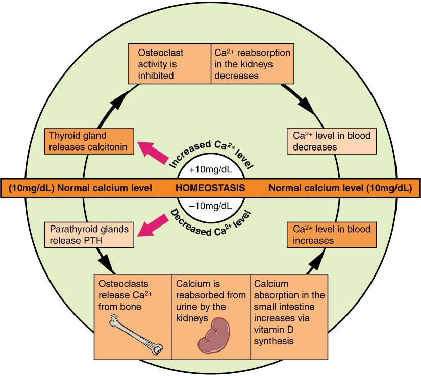
Three hormones defend the ionized calcium set-point: PTH (raises Ca; the fast, minute-to-minute regulator), calcitriol / 1,25-(OH)₂ vitamin D (raises Ca and phosphate; the gut absorber), and calcitonin (lowers Ca; minor in humans). FGF-23 is a fourth player that primarily governs phosphate.
- Chief cells of the parathyroid glands release PTH when the calcium-sensing receptor (CaSR) — a Gq-coupled GPCR — detects low ionized Ca²⁺. High Ca activates CaSR → suppresses PTH release.
- PTH acts on target cells through Gs → cAMP (the basis of urinary cAMP measurement).
- Bone: PTH binds osteoblasts, driving ↑ RANKL and ↓ osteoprotegerin (OPG) → osteoclast activation and resorption (osteoclasts have no PTH receptor — they are recruited indirectly). Ca and phosphate are liberated.
- Kidney (net: keep Ca, dump phosphate):
- ↑ Ca²⁺ reabsorption in the distal convoluted tubule (DCT)
- ↓ phosphate reabsorption in the proximal tubule (inhibits the Na⁺–phosphate cotransporter NaPi-IIa) → phosphaturia
- ↑ 1α-hydroxylase → more active vitamin D
- Net serum effect: ↑ calcium, ↓ phosphate.
- Magnesium twist: mild hypomagnesemia stimulates PTH, but severe hypomagnesemia inhibits PTH secretion AND causes PTH resistance → hypocalcemia that stays refractory until Mg²⁺ is repleted (think alcoholics, PPIs, loop/thiazide diuretics, diarrhea).

Hormone effects at a glance
| Hormone | Source & trigger | Gut | Bone | Kidney | Net serum |
|---|---|---|---|---|---|
| PTH | Chief cells; ↑ by ↓ ionized Ca | ↑ Ca (indirect, via calcitriol) | ↑ resorption (RANKL on osteoblasts) | ↑ Ca reabs (DCT); ↓ PO₄ reabs (PCT); ↑ 1α-hydroxylase | ↑ Ca, ↓ PO₄ |
| Calcitriol (1,25-D) | Kidney PCT; ↑ by ↑PTH, ↓PO₄, ↓Ca | ↑↑ Ca and PO₄ absorption (calbindin, TRPV6) | ↑ resorption (permissive with PTH) | ↑ Ca & PO₄ reabsorption (minor) | ↑ Ca, ↑ PO₄ |
| Calcitonin | Thyroid parafollicular C cells; ↑ by ↑Ca | — | ↓ osteoclast resorption | ↑ Ca & PO₄ excretion | ↓ Ca (weak; minor in humans) |
| FGF-23 | Osteocytes; ↑ by ↑PO₄, ↑calcitriol | ↓ PO₄ absorption (via ↓ calcitriol) | (site of secretion) | ↓ PO₄ reabsorption; ↓ 1α-hydroxylase, ↑ 24-hydroxylase | ↓ PO₄, ↓ calcitriol |
Vitamin D — two hydroxylations to activation
Vitamin D is a prohormone requiring two sequential hydroxylations:
- Skin: UVB light converts 7-dehydrocholesterol → cholecalciferol (D₃). Diet contributes D₃ (animal) or D₂ / ergocalciferol (plants, fortified foods).
- Liver: 25-hydroxylase → 25-OH-D (calcidiol) — the storage form and the best laboratory marker of vitamin D status. This step is essentially unregulated.
- Kidney (proximal tubule): 1α-hydroxylase → 1,25-(OH)₂-D (calcitriol) — the active hormone. This is the rate-limiting, tightly regulated step.
Regulation of 1α-hydroxylase:
- Stimulated by ↑ PTH, ↓ phosphate, ↓ calcium
- Inhibited by FGF-23, ↑ phosphate, and calcitriol itself (negative feedback; excess substrate is shunted to the inactive 24,25-(OH)₂-D).
Calcitriol's headline job is ↑ intestinal absorption of both calcium and phosphate — it is the only hormone that meaningfully increases gut calcium uptake.
- 25-OH-D = status marker; 1,25-(OH)₂-D = active hormone. Order 25-OH-D to diagnose deficiency; don't confuse the two.
- Calcitonin "tones down" calcium by inhibiting osteoclasts, but it is physiologically minor in humans: total thyroidectomy does not cause hypercalcemia, and the sky-high calcitonin of medullary thyroid carcinoma does not cause hypocalcemia (it's just a tumor marker there).
- FGF-23 = phosphaturic + anti-calcitriol, and requires the Klotho co-receptor. Excess FGF-23 → hypophosphatemia + rickets/osteomalacia (X-linked hypophosphatemic rickets = PHEX mutation; tumor-induced osteomalacia).
- Granulomatous disease and lymphoma carry unregulated 1α-hydroxylase in macrophages → autonomous calcitriol → hypercalcemia with a LOW PTH (classic sarcoidosis).
- ECG/signs: hypercalcemia shortens QT; hypocalcemia prolongs QT and produces Chvostek (cheek tap → facial twitch) and Trousseau (BP cuff → carpal spasm) signs.
Primary hyperparathyroidism. A 55-year-old woman with fatigue, constipation, and a kidney stone; Ca 11.2, phosphate low, PTH high. Mechanism: an autonomous parathyroid adenoma. Chronic resorption produces osteitis fibrosa cystica (subperiosteal resorption, brown tumors), a raised alkaline phosphatase, and hypercalciuria → stones — "stones, bones, groans, and psychiatric overtones."
Familial hypocalciuric hypercalcemia (FHH). Asymptomatic hypercalcemia with an inappropriately normal/high PTH but a LOW urine calcium. Mechanism: an inactivating CaSR mutation raises the calcium set-point and drives renal Ca reabsorption. Benign — do not operate; the low urine Ca distinguishes it from primary hyperparathyroidism (which spills high urine Ca).
Hypoparathyroidism. Perioral numbness and carpopedal spasm after total thyroidectomy (surgery is the #1 cause), or DiGeorge (22q11.2 deletion). Labs: low PTH, low Ca, HIGH phosphate, prolonged QT.
Pseudohypoparathyroidism type 1A. Low Ca, high phosphate, but PTH is HIGH — end-organ resistance from a defective Gsα protein (GNAS, maternally inherited). Phenotype = Albright hereditary osteodystrophy: short stature, round face, and shortened 4th/5th metacarpals.
Humoral hypercalcemia of malignancy. Squamous cell carcinoma (lung, head & neck) or renal cell carcinoma secretes PTHrP → high Ca, low phosphate, but PTH is LOW (suppressed) and 1,25-D is low/normal — the opposite pattern to primary hyperparathyroidism.
Secondary hyperparathyroidism of CKD. The failing kidney can't make calcitriol and retains phosphate → serum Ca falls → PTH climbs; here phosphate is HIGH (contrast nutritional vitamin D deficiency, where phosphate is low). Long-standing stimulation can become autonomous — tertiary hyperparathyroidism, now with high Ca.

Lab pattern differential (the money table)
| Disorder | Ca | PO₄ | PTH | Discriminating clue |
|---|---|---|---|---|
| Primary hyperPTH (adenoma) | ↑ | ↓ | ↑ | ↑ urine Ca, ↑ 1,25-D |
| FHH | ↑ | nl | nl/↑ | ↓ urine Ca (don't operate) |
| Malignancy — PTHrP | ↑ | ↓ | ↓ | squamous / renal cell cancer |
| Sarcoidosis / lymphoma | ↑ | nl/↑ | ↓ | ↑ 1,25-D (macrophage 1α-hydroxylase) |
| Hypoparathyroidism | ↓ | ↑ | ↓ | post-thyroidectomy / DiGeorge |
| Pseudohypoparathyroidism 1A | ↓ | ↑ | ↑ | Gsα resistance + Albright phenotype |
| Vitamin D deficiency (rickets/osteomalacia) | ↓/nl | ↓ | ↑ | ↓ 25-OH-D, ↑ alk phos |
| CKD (secondary hyperPTH) | ↓/nl | ↑ | ↑ | ↓ 1,25-D, renal osteodystrophy |
- Calci-TONE-in TONES DOWN blood calcium (inhibits osteoclasts).
- Hypercalcemia symptoms: "Stones, Bones, Groans, Thrones, and Psychiatric Overtones" — kidney stones, bone pain, abdominal/GI groans (constipation, PUD, pancreatitis), thrones = polyuria, and depression/confusion.
- PTH = "Phosphate Trashing Hormone" — it drives phosphaturia, so PTH raises Ca but lowers serum phosphate.
- Chvostek = Cheek (tap the cheek → facial twitch); Trousseau = Tourniquet (inflate the BP cuff → carpal spasm) — both signs of hypocalcemia.
- Vitamin D activation route: liver adds the 25, kidney adds the 1 (2 hydroxylations; the kidney's 1α-hydroxylase is the regulated, active-forming step).
Practice Physiology now
Board-style questions, spaced-repetition flashcards, and a Socratic AI tutor — free to start.