Calcium & Phosphate Disorders
Calcium and phosphate are governed by PTH, vitamin D, and FGF23 across gut, bone, and kidney — and boards test the lab pattern (Ca / PO₄ / PTH) more than any single value. This lesson walks the PTH-based workup of hyper- and hypocalcemia, the CHIMPANZEES causes, next-best-step management, and the phosphate/CKD-MBD picture.
The Calcium–Phosphate Axis
Serum calcium and phosphate are held in balance by three hormones acting on gut, bone, and kidney. Boards test the pattern they produce far more than any single number.
- PTH (parathyroid, sensed by the CaSR) is released when Ca falls: it raises Ca and lowers phosphate — resorbs bone, reabsorbs Ca in the distal tubule, wastes phosphate proximally, and switches on renal 1α-hydroxylase.
- Calcitriol (1,25-(OH)₂ vitamin D) raises both Ca and phosphate by boosting gut absorption of each.
- FGF23 (from osteocytes) lowers phosphate (phosphaturia) and suppresses calcitriol — the key driver of CKD mineral disease.
- Calcitonin (thyroid C cells) modestly lowers Ca; minor in humans.
About 50% of serum Ca is the biologically active ionized fraction; roughly 40% is albumin-bound and ~10% complexed with anions. Always correct for albumin: corrected Ca = measured Ca + 0.8 × (4.0 − albumin). In critical illness or citrate load, measure ionized calcium directly — alkalosis drops ionized Ca (perioral tingling in the hyperventilating patient).
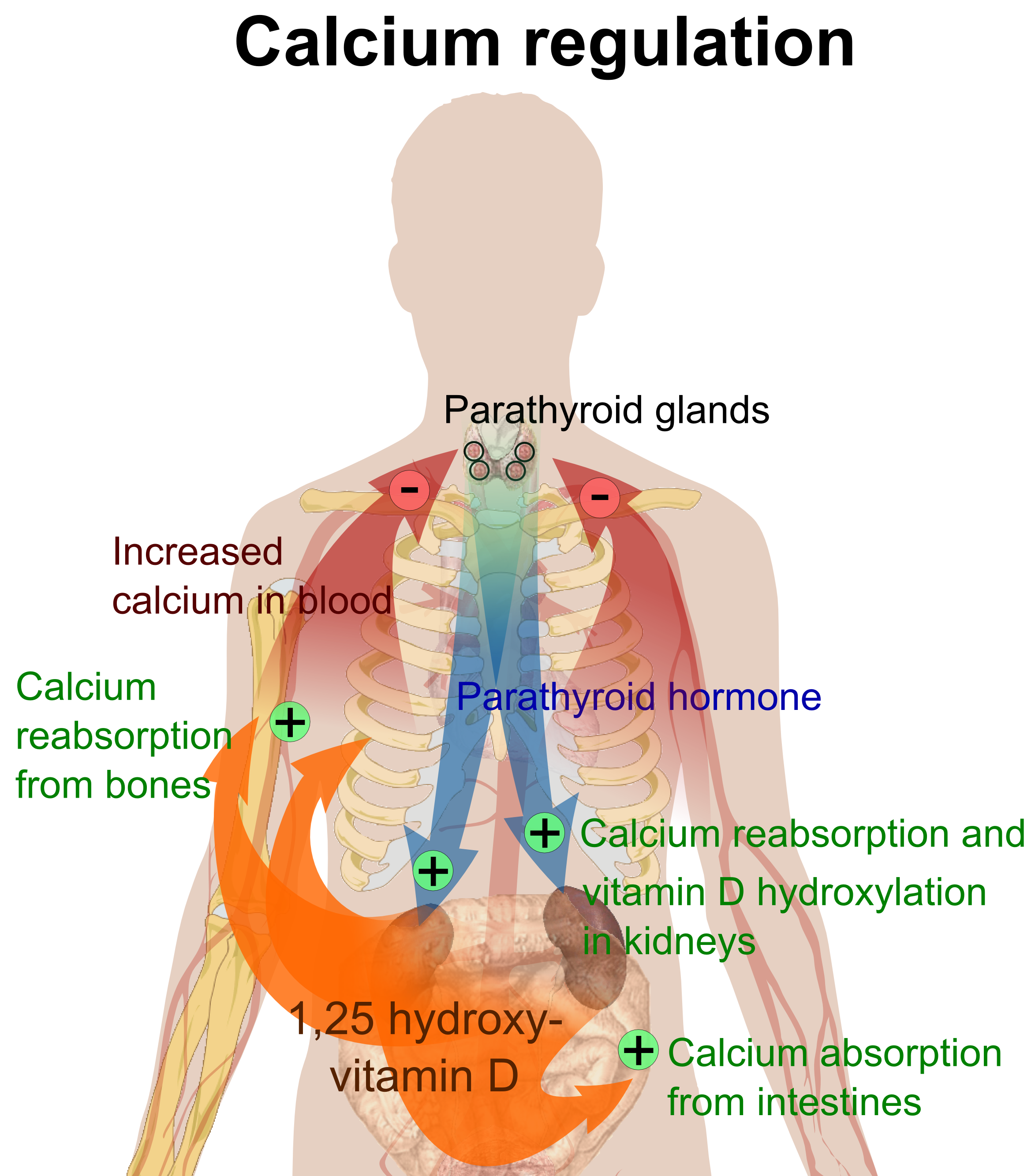
- Normal total Ca 8.5–10.5 mg/dL · phosphate 2.5–4.5 mg/dL
- Corrected Ca = Ca + 0.8 × (4.0 − albumin g/dL) — low albumin fakes low total Ca (ionized is normal)
- PTH → ↑Ca, ↓PO₄ · Vitamin D → ↑Ca, ↑PO₄ · FGF23 → ↓PO₄
- Symptomatic hypercalcemia usually > 12 mg/dL; > 14 or symptomatic = emergency
- ECG: hypercalcemia → short QT; hypocalcemia → long QT (torsades risk)
- ~90% of hypercalcemia = primary hyperparathyroidism (outpatient, mild) or malignancy (inpatient, sicker, higher Ca)
- First test in hypercalcemia = PTH — it splits PTH-dependent vs PTH-independent
- Hypomagnesemia both blocks PTH release and causes PTH resistance → refractory hypocalcemia; replace Mg first
Reading the Labs: Classic Patterns
| Disorder | Ca | PO₄ | PTH | Clue |
|---|---|---|---|---|
| 1° hyperparathyroidism | ↑ | ↓ | ↑ | Adenoma; ↑ urine Ca; incidental ↑Ca |
| Malignancy (PTHrP) | ↑↑ | ↓/↔ | ↓ | Squamous lung, renal, breast; sick |
| FHH | ↑ | ↔ | ↔/↑ | Low urine Ca (Ca/Cr clearance <0.01); CaSR |
| Vitamin D deficiency | ↓/↔ | ↓ | ↑ | ↓ 25-OH D; 2° hyperPTH |
| Hypoparathyroidism | ↓ | ↑ | ↓ | Post-thyroidectomy |
| Pseudohypoparathyroidism | ↓ | ↑ | ↑ | PTH resistance (Albright osteodystrophy) |
| CKD (2° hyperPTH) | ↓/↔ | ↑ | ↑ | ↑ phosphate, ↓ calcitriol, ↑ FGF23 |
Causes of hypercalcemia (CHIMPANZEES):
- C — Calcium supplementation
- H — Hyperparathyroidism / Hyperthyroidism
- I — Iatrogenic (thiazides) / Immobilization
- M — Milk-alkali / Multiple myeloma
- P — Paget disease / Parenteral nutrition
- A — Addison disease / Acromegaly
- N — Neoplasm (PTHrP, osteolytic mets)
- Z — Zollinger-Ellison (as part of MEN1)
- E — Excess vitamin D
- E — Excess vitamin A
- S — Sarcoidosis / granulomatous disease
Symptoms — 'stones, bones, groans, thrones, psychiatric overtones': kidney stones, bone pain, abdominal groans (constipation, PUD, pancreatitis), polyuria (thrones), and confusion/depression.
Vignette: An asymptomatic 58-year-old woman has Ca 11.2, PO₄ 2.2 on routine labs.
- Next step = check PTH. Here PTH is high-normal/elevated → PTH-dependent. High Ca + low phosphate + high PTH = primary hyperparathyroidism (adenoma).
- Before parathyroidectomy, exclude FHH: order a 24-h urine calcium / Ca-creatinine clearance ratio. Low urine Ca (<0.01) → FHH — do NOT operate (benign, autosomal-dominant CaSR defect).
- Low/suppressed PTH instead → PTH-independent → hunt malignancy: check PTHrP (squamous lung, renal, breast, bladder), 1,25-vitamin D (lymphoma, sarcoidosis), and SPEP/imaging for myeloma / osteolytic mets.
Buzzwords: band keratopathy, subperiosteal resorption (radial side of the middle phalanges), osteitis fibrosa cystica with brown tumors, 'salt-and-pepper' skull, short QT.

Management — Emergencies Both Ways
Acute severe hypercalcemia (Ca > 14 or symptomatic):
- Aggressive IV isotonic saline — restores volume and drives calciuresis (dehydration is universal). Always first.
- Calcitonin — works within hours but tachyphylaxis by ~48 h; a bridge only.
- IV bisphosphonate (zoledronic acid) — the durable fix; onset 2–4 days. Denosumab if refractory or renal failure.
- Glucocorticoids for vitamin-D-mediated causes (lymphoma, sarcoidosis, vitamin D toxicity).
Loop diuretics are no longer routine — reserve for volume overload; hemodialysis for renal failure/refractory cases.
Symptomatic hypocalcemia (tetany, seizure, laryngospasm, long QT): IV calcium gluconate. Chronic: oral calcium + activated vitamin D (calcitriol). Always check and replace magnesium — hypomagnesemia causes PTH-resistant hypocalcemia that won't correct until Mg is restored.
Vignette: 12 hours after a total thyroidectomy, a patient reports perioral numbness and finger tingling; the BP cuff triggers carpal spasm.
- Diagnosis: acute hypocalcemia from inadvertent parathyroid removal/injury — postsurgical hypoparathyroidism, the classic cause. Labs: low Ca, high phosphate, low PTH.
- Bedside signs: Trousseau sign (carpopedal spasm on BP-cuff inflation — more specific) and Chvostek sign (facial twitch on tapping CN VII).
- Next step: confirm with ionized Ca, obtain an ECG (prolonged QT), and give IV calcium gluconate for symptomatic tetany.
Other tested triggers: acute pancreatitis (saponification), tumor lysis / rhabdomyolysis (hyperphosphatemia), massive transfusion (citrate), respiratory alkalosis (↓ ionized Ca — the panicked hyperventilator), and hypomagnesemia (alcoholic, PPI use).

Phosphate & CKD–Mineral Bone Disorder
Hyperphosphatemia — most often CKD (can't excrete), plus tumor lysis, rhabdomyolysis, and hypoparathyroidism. It binds calcium → hypocalcemia and drives PTH. Treat with dietary restriction and phosphate binders (sevelamer, calcium acetate; avoid long-term aluminum).
Hypophosphatemia — board classics: refeeding syndrome, DKA treatment (insulin shifts PO₄ into cells), alcoholism, and respiratory alkalosis. Severe (< 1 mg/dL) → rhabdomyolysis, hemolysis, and respiratory-muscle weakness. FGF23 excess wastes phosphate renally (X-linked hypophosphatemic rickets [PHEX], oncogenic osteomalacia).
CKD-MBD: failing kidneys retain phosphate and can't make calcitriol → hypocalcemia with rising FGF23 → secondary hyperparathyroidism → renal osteodystrophy. Untreated, the glands go autonomous — tertiary hyperparathyroidism with hypercalcemia and very high PTH.
Hyper- vs Hypocalcemia at a Glance
| Feature | Hypercalcemia | Hypocalcemia |
|---|---|---|
| Neuro/psych | Lethargy, confusion, weakness, coma | Tetany, seizures, irritability, paresthesias |
| Classic signs | Stones, bones, groans, thrones | Chvostek & Trousseau, perioral numbness |
| GI | Constipation, PUD, pancreatitis | Cramps |
| Cardiac / ECG | Short QT, hypertension | Long QT (torsades), laryngospasm |
| Renal | Nephrolithiasis, nephrogenic DI (polyuria) | — |
| Emergency Rx | IV saline → calcitonin → bisphosphonate | IV calcium gluconate (+ replace Mg) |
Practice Renal now
Board-style questions, spaced-repetition flashcards, and a Socratic AI tutor — free to start.