Brachial Plexus & Upper-Limb Nerve Injuries
The brachial plexus (C5–T1) and the five terminal nerves it spawns, mapped to the fractures, dislocations, and birth injuries that classically damage them — with the wrist-drop, ape-hand, claw-hand, benediction, and waiter's-tip signs that boards love to test, and the proximal-vs-distal lesion levels that change the picture.
Organization: roots to branches
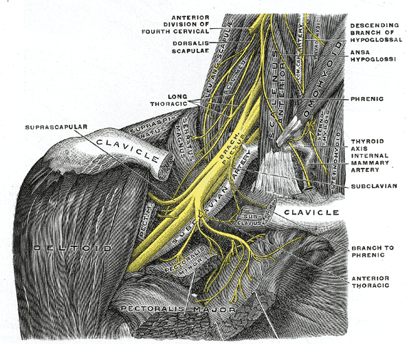
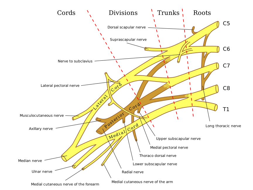
The brachial plexus supplies all motor and sensory innervation to the upper limb, arising from the ventral rami of C5–T1. Fibers reorganize through five stages, proximal to distal — Roots → Trunks → Divisions → Cords → Branches:
- Trunks: upper (C5–C6), middle (C7), lower (C8–T1).
- Cords are named for their position around the second part of the axillary artery: lateral, posterior, medial.
- Every posterior division feeds the posterior cord, which gives off the axillary and radial nerves — the limb's abductors and extensors. The lateral and medial cords each send a root that fuses to form the median nerve.
The test rarely asks you to redraw the plexus — it drops a fracture or dislocation and expects you to name the injured nerve and its deficit.
- "Randy Travis Drinks Cold Beer" = Roots → Trunks → Divisions → Cords → Branches (proximal to distal).
- The "M" sign: overlying the axillary artery, the musculocutaneous nerve, the median nerve, and the ulnar nerve — linked by the lateral and medial cords — trace a capital M. The median nerve sits at the central junction of the M, formed where its lateral and medial roots meet.
- Axillary (C5–C6): surgical neck of humerus fracture / anterior shoulder dislocation → deltoid + teres minor palsy (weak abduction 15–90°), sensory loss over the lateral shoulder ("regimental badge").
- Radial (C5–T1): midshaft humerus fracture or spiral-groove compression ("Saturday-night palsy" — arm draped over a chair back) → wrist drop, lost wrist/finger/thumb extension; sensory loss over dorsal hand + first web space. Triceps is spared at the spiral groove (its branches leave proximally) — but axillary "crutch palsy" also weakens the triceps.
- Median — proximal (supracondylar humerus fracture): lost forearm pronation and flexion of digits 2–3 → "hand of benediction" when making a fist, plus thenar atrophy ("ape hand") and lost opposition.
- Median — distal (carpal tunnel): thenar atrophy / ape hand + lost opposition only — forearm long flexors are spared, so no benediction; sensory loss over palmar lateral 3½ digits (central palm spared by the palmar cutaneous branch).
- Ulnar (C8–T1): medial epicondyle ("funny bone") or hook-of-hamate fracture → "ulnar claw," lost finger abduction/adduction, positive Froment sign; sensory loss over medial 1½ digits.
- Musculocutaneous (C5–C7): lost elbow flexion + supination; sensory loss over the lateral forearm.
- Long thoracic (C5–C7): serratus anterior palsy → winged scapula (classic after axillary node dissection / mastectomy).
The five terminal nerves at a glance
| Nerve | Roots | Classic injury | Motor deficit | Sensory loss |
|---|---|---|---|---|
| Axillary | C5–C6 | Surgical neck fx / ant. dislocation | Deltoid, teres minor (abduction 15–90°) | Lateral shoulder (regimental badge) |
| Musculocutaneous | C5–C7 | Upper trunk / direct trauma | Biceps, brachialis (elbow flexion + supination) | Lateral forearm |
| Radial | C5–T1 | Midshaft humerus / spiral groove | Wrist + finger extensors → wrist drop | Dorsal hand, first web space |
| Median | C5–T1 | Supracondylar fx (proximal) / carpal tunnel (distal) | Thenar + opposition; digit 2–3 flexion only if proximal | Palmar lateral 3½ digits |
| Ulnar | C8–T1 | Medial epicondyle / hook of hamate | Interossei, medial lumbricals, adductor pollicis | Medial 1½ digits |
The vignette hands you a bone and expects the nerve:
- Surgical neck of humerus / anterior dislocation → axillary.
- Midshaft humerus → radial (wrist drop).
- Supracondylar humerus → median (watch for the brachial artery too).
- Medial epicondyle → ulnar.
- Hook of hamate (fall on outstretched hand, cyclist gripping handlebars) → distal ulnar (Guyon canal).
- Fall on the shoulder / birth shoulder dystocia → upper trunk (Erb).
- Upward traction on an abducted arm → lower trunk (Klumpke); look for Horner syndrome if T1 is involved.
Buzzword-to-next-step: "fell asleep with the arm draped over a chair" + wrist drop → radial (spiral-groove) compression; nocturnal tingling + thenar wasting → carpal tunnel → confirm with Phalen / Tinel.
Erb–Duchenne vs Klumpke
| Feature | Erb–Duchenne | Klumpke |
|---|---|---|
| Lesion | Upper trunk C5–C6 | Lower trunk C8–T1 |
| Mechanism | Downward shoulder traction (shoulder dystocia, fall on shoulder, motorcycle) | Upward traction on an abducted arm (breech grab, catching a branch mid-fall) |
| Deficit | Deltoid, biceps, rotator cuff | Intrinsic hand muscles |
| Posture | "Waiter's tip" — arm adducted, internally rotated, elbow extended, forearm pronated | Total claw hand |
| Extra clue | — | Horner syndrome (ptosis, miosis, anhidrosis) if T1 sympathetics involved |
Claw vs benediction — and the ulnar paradox
These deformities look alike but are triggered differently — and the level of the lesion changes the picture:
- "Hand of benediction" = a proximal median lesion (e.g., supracondylar fracture), revealed when the patient tries to make a fist — digits 2–3 stay extended because flexion of the lateral fingers (FDS, FDP to 2–3, FPL) is lost. A distal median lesion (carpal tunnel) spares those forearm flexors, so it gives thenar wasting / ape hand without benediction.
- "Ulnar claw" = an ulnar lesion, seen at rest — the ring and little fingers hyperextend at the MCP and flex at the IP joints (unopposed extensors + weak medial lumbricals).
- Ulnar paradox: a distal ulnar lesion produces a worse-looking claw than a proximal one. A proximal (elbow) lesion also denervates FDP to digits 4–5, so those DIPs can't flex tightly — the more severe, proximal injury paradoxically looks less clawed.
- Mirror image: the median benediction leaves digits 2–3 extended; the ulnar claw involves digits 4–5.
- Abduction split: supraspinatus initiates the first ~15°; the deltoid (axillary) drives 15–90° — so an axillary lesion still permits a little abduction.
- Anterior interosseous nerve (pure-motor median branch): can't make the "OK" sign (weak FPL + FDP to index), with no sensory loss.
- Posterior interosseous nerve (deep radial branch): finger drop with preserved wrist extension (wrist deviates radially) and no sensory loss.
- Froment sign (ulnar): the thumb IP flexes (FPL substitutes) when pinching paper because adductor pollicis is weak.
- T1 deficit + Horner syndrome → think lower-trunk lesion or a Pancoast (apical lung) tumor.
Practice Anatomy now
Board-style questions, spaced-repetition flashcards, and a Socratic AI tutor — free to start.