Blood & Bone Marrow Histology
A board-focused tour of the peripheral smear and bone marrow: identifying each formed element, the leukocyte-frequency mnemonic, and the classic morphologic buzzwords (hypersegmented neutrophils, Auer rods, schistocytes) that map to Step 1 diagnoses and next-best-steps.
Overview: the smear and the marrow
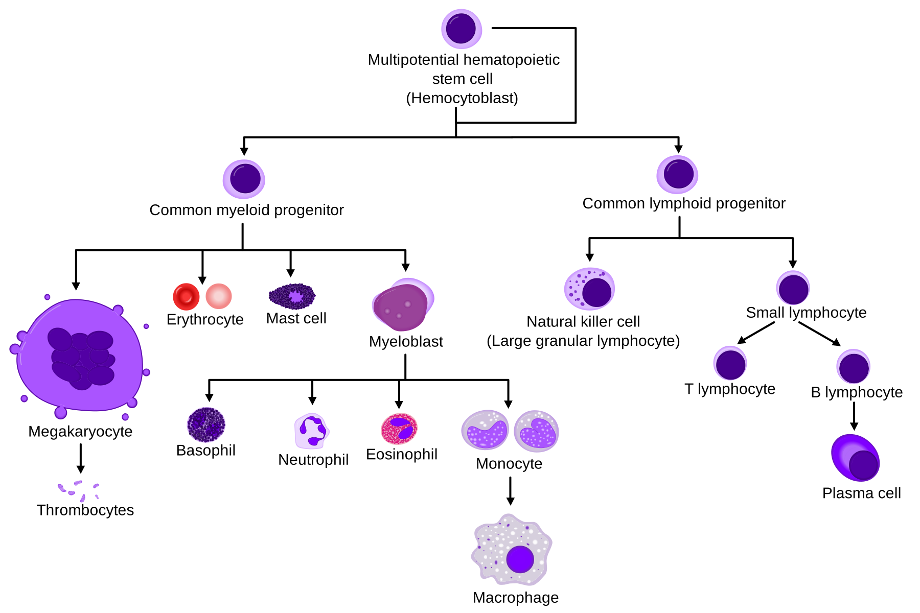
Blood is a specialized connective tissue: formed elements (RBCs, WBCs, platelets) suspended in plasma. On a Wright-Giemsa stained peripheral smear, the RBC (7.5 μm, central pallor) is your internal ruler for sizing every other cell. Adult hematopoiesis occurs in red bone marrow of the axial skeleton (vertebrae, sternum, ribs, iliac crest) plus the proximal epiphyses of the femur and humerus. All lineages arise from one pluripotent hematopoietic stem cell (HSC) that splits into common myeloid and common lymphoid progenitors. Fetal hematopoiesis follows Yolk sac → Liver → Spleen → Bone marrow, with the liver dominant in the 2nd trimester and marrow taking over by birth. Boards test three things: (1) morphology recognition — which cell is this?, (2) the buzzword inclusion on the smear that points to a diagnosis, and (3) the classic leukocyte-frequency mnemonic.
- Erythrocyte: 7.5 μm biconcave disc, anucleate, no organelles, ~120-day lifespan; central pallor ≈ 1/3 of the diameter
- Reticulocyte: young RBC with residual ribosomal RNA; shows polychromasia (bluish) on Wright stain and a reticular mesh on supravital stain (new methylene blue); normal ~0.5–1.5%
- Reticulocytosis = appropriate marrow response to anemia (hemolysis or blood loss); low retic = production problem
- Platelet (thrombocyte): 2–3 μm anucleate cytoplasmic fragment shed from a megakaryocyte; ~8–10-day lifespan
- Size cues: microcytic RBC < small lymphocyte nucleus; macrocytic RBC > that nucleus
- Anisocytosis = variable size (↑RDW); poikilocytosis = variable shape
The five leukocytes
| WBC | Nucleus | Granules / cytoplasm | Role / board buzzword |
|---|---|---|---|
| Neutrophil | 3–5 lobes | fine lilac-pink | Acute bacterial; hypersegmented = B12/folate; left shift = bands |
| Eosinophil | bilobed | large red-orange | Parasites, type I allergy; NAACP causes |
| Basophil | bilobed (obscured) | large dark blue-purple | Histamine/heparin; basophilia → CML |
| Lymphocyte | round, dense, ~cell-sized | thin blue rim | B/T/NK; reactive in viral (mono) |
| Monocyte | kidney / horseshoe | abundant gray-blue, vacuoles | Largest WBC; → macrophage/dendritic |
- Frequency (high → low): "Never Let Monkeys Eat Bananas" = Neutrophils > Lymphocytes > Monocytes > Eosinophils > Basophils
- Approximate differential 60 / 30 / 6 / 3 / 1 percent — neutrophils dominate, basophils are rarest (<1%)
- Eosinophilia — "NAACP": Neoplasia, Allergy/Asthma, Addison disease, Collagen-vascular disease, Parasites
- Left shift = immature bands and precursors released from marrow → bacterial infection or marrow stress
Vignette: A 68-year-old strict vegan reports fatigue and tingling in the feet. Hb 9.1, MCV 116. Smear shows macro-ovalocytes and hypersegmented neutrophils (>5 lobes).
- Diagnosis: megaloblastic anemia (B12 or folate deficiency) — impaired DNA synthesis causes nuclear–cytoplasmic asynchrony.
- Next best step: measure serum B12 and folate; if equivocal, check methylmalonic acid (MMA) + homocysteine.
- ↑MMA and ↑homocysteine → B12 deficiency
- normal MMA, ↑homocysteine → folate deficiency
- Neuro signs (subacute combined degeneration of dorsal columns/lateral corticospinal tract) indicate B12, not folate. Replace B12 before/with folate so folate doesn't correct the anemia while neuro damage progresses.
- Red (active) marrow in adults: axial skeleton + proximal epiphyses of femur/humerus; yellow (fatty) marrow elsewhere and reconverts under high demand
- At birth all marrow is red; fat progressively replaces it with age
- Myeloid:erythroid (M:E) ratio normally ~2:1 to 3:1 (granulocyte precursors outnumber erythroid)
- Megakaryocytes: the largest marrow cells — huge lobulated polyploid nucleus; extend proplatelet processes to shed platelets into marrow sinusoids
- Preferred biopsy site: posterior iliac crest
- >20% blasts = acute leukemia; normal cellularity ≈ (100 − age)% — hypercellular in leukemia, hypocellular in aplastic anemia
Vignette: A 62-year-old presents with fatigue, gum bleeding, and pancytopenia except WBC 40,000. Marrow shows 30% myeloblasts; several blasts contain pink, needle-shaped Auer rods.
- Diagnosis: Acute myeloid leukemia (AML) — Auer rods are peroxidase (MPO)-positive fused azurophilic granules; >20% blasts confirms acute leukemia.
- Buzzword branch: APL (AML-M3, t(15;17), PML-RARA) shows abundant Auer rods (faggot cells) and high DIC risk; treat with all-trans retinoic acid (ATRA).
- Contrast: smudge cells → CLL; >20% blasts + Auer rods → AML; t(9;22) Philadelphia with basophilia → CML.
- Next step: flow cytometry + cytogenetics for classification and prognosis.
Smear buzzwords → diagnosis
| Smear finding | Classic association |
|---|---|
| Hypersegmented neutrophils | B12 / folate (megaloblastic) |
| Howell-Jolly bodies | Asplenia (nuclear remnants) |
| Basophilic stippling | Lead poisoning, sideroblastic anemia, thalassemia |
| Target cells | HbC, Asplenia, Liver disease, Thalassemia ("HALT") |
| Schistocytes | MAHA — DIC, TTP/HUS, mechanical valve |
| Bite cells / Heinz bodies | G6PD deficiency |
| Spherocytes | Hereditary spherocytosis, warm AIHA |
| Teardrop cells (dacrocytes) | Myelofibrosis |
| Rouleaux | Multiple myeloma |
Practice Histology now
Board-style questions, spaced-repetition flashcards, and a Socratic AI tutor — free to start.