Bleeding Disorders: Hemophilia & von Willebrand
A high-yield STEP 1 review contrasting hemophilia A/B (factor VIII/IX deficiency, X-linked recessive, deep bleeding, isolated prolonged PTT that corrects on mixing) with von Willebrand disease (most common inherited bleeding disorder, mucocutaneous bleeding, prolonged bleeding time/PFA). It drills the lab reflexes, ristocetin logic, DDAVP-vs-concentrate decisions, inhibitor pitfalls, and classic next-best-step vignettes. Corrected the DDAVP mechanism: Weibel–Palade bodies store vWF (not factor VIII); DDAVP raises factor VIII indirectly via released vWF.
Two flavors of bleeding
Bleeding disorders split into platelet-type (mucocutaneous: epistaxis, gum bleeding, petechiae, menorrhagia, immediate post-op oozing) and coagulation-factor-type (deep: hemarthrosis, muscle hematomas, delayed rebleeding after trauma/surgery). Hemophilia A (factor VIII) and B (factor IX) are the classic factor-deficiency diseases — X-linked recessive, so nearly all patients are male. von Willebrand disease (vWD) is the most common inherited bleeding disorder (usually autosomal dominant) and behaves like a platelet problem because vWF anchors platelets to collagen.
Boards hinge on one reflex: an isolated prolonged PTT with normal PT points to the intrinsic pathway (VIII, IX) → hemophilia; add a prolonged bleeding time / abnormal PFA and you have vWD. Everything else — which factor, DDAVP vs concentrate, the ristocetin test — flows from that single lab pattern.
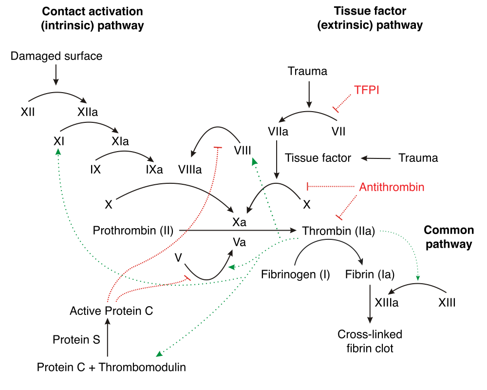
- Hemophilia A = factor VIII deficiency (most common); Hemophilia B ("Christmas disease") = factor IX. Clinically identical.
- Both X-linked recessive → males affected, females carriers; up to 1/3 are spontaneous mutations (no family history).
- Labs: ↑PTT, normal PT, normal platelet count, normal bleeding time. Severity tracks factor activity (<1% = severe, spontaneous hemarthrosis).
- Mixing (1:1) study corrects in a factor deficiency; fails to correct = inhibitor (alloantibody in treated hemophiliacs, or acquired hemophilia A in elderly/postpartum).
- vWD: ↓vWF → defective platelet adhesion and low factor VIII (vWF stabilizes VIII) → prolonged bleeding time/PFA, PTT normal or mildly prolonged, normal PT.
- vWF has two jobs: bridges platelet GpIb to subendothelial collagen, and carries/protects factor VIII.
- Mucocutaneous bleeding + normal platelet count + prolonged bleeding time → think vWD first (most common).

Lab & marker comparison
| Feature | Hemophilia A / B | von Willebrand disease |
|---|---|---|
| Inheritance | X-linked recessive | AD (type 3 = AR) |
| Bleeding pattern | Deep — hemarthrosis, hematomas | Mucocutaneous — epistaxis, menorrhagia |
| Platelet count | Normal | Normal (↓ in type 2B) |
| PT | Normal | Normal |
| PTT | Prolonged | Normal or mildly ↑ |
| Bleeding time / PFA | Normal | Prolonged |
| Factor VIII | Low (A) / normal (B) | Low–normal |
| vWF antigen | Normal | Decreased |
| Ristocetin cofactor | Normal | Decreased |
| Peripheral smear | Normal | Normal |
| First-line Rx | Factor concentrate; DDAVP (mild A) | DDAVP; vWF concentrate |
A 5-year-old boy has a warm, swollen, painful knee after minor play, plus a history of prolonged bleeding after circumcision and easy deep bruising. A maternal uncle had similar problems. Labs: platelets normal, PT normal, PTT prolonged, and the PTT corrects on a 1:1 mix with normal plasma.
- Diagnosis: Hemophilia (A or B) — confirm with factor VIII and factor IX activity assays.
- Why: X-linked pattern (maternal uncle), deep/joint bleeding, isolated ↑PTT that corrects on mixing (a deficiency, not an inhibitor). Peripheral smear adds nothing here.
- Next best step (acute bleed): replace the deficient factor — recombinant factor VIII (hemophilia A) or factor IX (B). DDAVP works only for mild hemophilia A.
- Prophylaxis pearl: emicizumab — a bispecific antibody bridging factor IXa and factor X (mimicking the cofactor role of VIIIa) — is used for hemophilia A, including patients with inhibitors.
A 16-year-old girl has lifelong menorrhagia, recurrent epistaxis, and prolonged bleeding after dental work. Labs: platelet count normal, PT normal, bleeding time (PFA) prolonged, PTT slightly prolonged.
- Diagnosis: von Willebrand disease — the most common inherited bleeding disorder.
- Confirm: ↓vWF antigen, ↓ristocetin cofactor activity, ↓factor VIII. Ristocetin-induced platelet agglutination is low and corrects when normal plasma (vWF) is added.
- Distinguish: in Bernard–Soulier (GpIb defect) ristocetin agglutination does NOT correct with normal plasma (plus large platelets, thrombocytopenia); Glanzmann thrombasthenia (GpIIb/IIIa defect) has normal ristocetin but absent aggregation to ADP/collagen/epinephrine.
- Next best step: DDAVP — releases vWF from endothelial Weibel–Palade bodies (best for type 1). Use vWF-containing concentrate for type 3 or severe type 2. Avoid DDAVP in type 2B (worsens thrombocytopenia).
- Hemophilia B = "Christmas disease" = factor IX (named for patient Stephen Christmas). Pair them: A ↔ VIII, B ↔ IX.
- PT → Extrinsic (factor VII); by elimination PTT → intrinsic pathway (XII, XI, IX, VIII). An isolated ↑PTT → suspect hemophilia.
- Ristocetin = the "vWF–GpIb test." It needs both vWF and platelet GpIb, so agglutination is poor in vWD (corrects with added plasma) and in Bernard–Soulier (does not correct).
- "vWF carries VIII": low vWF drops factor VIII, so severe vWD can also nudge the PTT upward.
- DDAVP (desmopressin) releases stored vWF from Weibel–Palade bodies and raises factor VIII levels (released vWF stabilizes and carries VIII, plus endothelial FVIII release) → works for mild hemophilia A and type 1 vWD. Useless in hemophilia B and severe/type 3 disease (no functional factor to boost).
- Contraindication: DDAVP in vWD type 2B worsens thrombocytopenia (mutant vWF binds platelets → clearance). Also watch hyponatremia (ADH/V2 analog) — restrict free water.
- Hemophilia A with rising bleeds despite replacement → suspect a factor VIII inhibitor: PTT mixing study won't correct; quantify with the Bethesda assay. Treat with bypassing agents (aPCC, recombinant factor VIIa) or emicizumab.
- Acquired hemophilia A: older adult or postpartum patient with new deep bleeding + isolated ↑PTT not correcting on mixing → autoantibody to factor VIII.
- All hemophilia: avoid aspirin/NSAIDs and IM injections; joint-directed prophylactic factor prevents arthropathy.
- Neonate, prolonged bleeding after circumcision, isolated ↑PTT → order factor VIII/IX activity assays (diagnose hemophilia), not a smear.
- Woman with menorrhagia + normal platelets + prolonged PFA/bleeding time → vWF antigen + ristocetin cofactor + factor VIII (vWD workup).
- Known mild hemophilia A, minor bleed → DDAVP first; major bleed or surgery → factor VIII concentrate dosed to target level.
- ↑PTT that does NOT correct on mixing → inhibitor (get a Bethesda titer), not a simple deficiency.
- Isolated ↑PTT but asymptomatic / no bleeding → classic distractor factor XII (Hageman) deficiency — prolongs PTT yet causes no bleeding.
Practice Heme/Onc now
Board-style questions, spaced-repetition flashcards, and a Socratic AI tutor — free to start.