Bacterial & Viral Skin Infections
A boards-focused tour of bacterial (impetigo, erysipelas, cellulitis, SSSS, necrotizing fasciitis) and viral (HSV, VZV, molluscum) skin infections, organized by tissue depth and buzzword morphology, with vignettes drilling next-best-step decisions and comparison tables for depth-based bacteria and SSSS vs. TEN.
Framework: Depth Predicts Organism and Severity
Skin infections are best organized by the layer involved, because depth predicts both morphology and the likely organism. Bacterial disease ascends a severity gradient: superficial epidermis (impetigo) → upper dermis and lymphatics (erysipelas) → deep dermis/subcutis (cellulitis) → fascia and muscle (necrotizing fasciitis). Two pathogens dominate: Staphylococcus aureus (including MRSA) and group A Streptococcus (S. pyogenes). Some "infections" are actually toxin-mediated — the toxin is produced at a distant focus (SSSS, toxic shock) — so blister or lesion cultures at the skin can be sterile.
Viral eruptions cluster into herpesviruses (HSV, VZV — grouped vesicles, multinucleated giant cells on Tzanck), poxvirus (molluscum), and HPV (warts). Boards reward pattern recognition: map the buzzword lesion (honey crust, dermatomal vesicles, umbilicated papule) to an organism, then pick the next best step — usually empiric therapy, sometimes a confirmatory test, occasionally urgent surgery. Always scan for red flags (pain out of proportion, crepitus, systemic toxicity) that convert a "prescribe antibiotics" item into a "call surgery now" item.
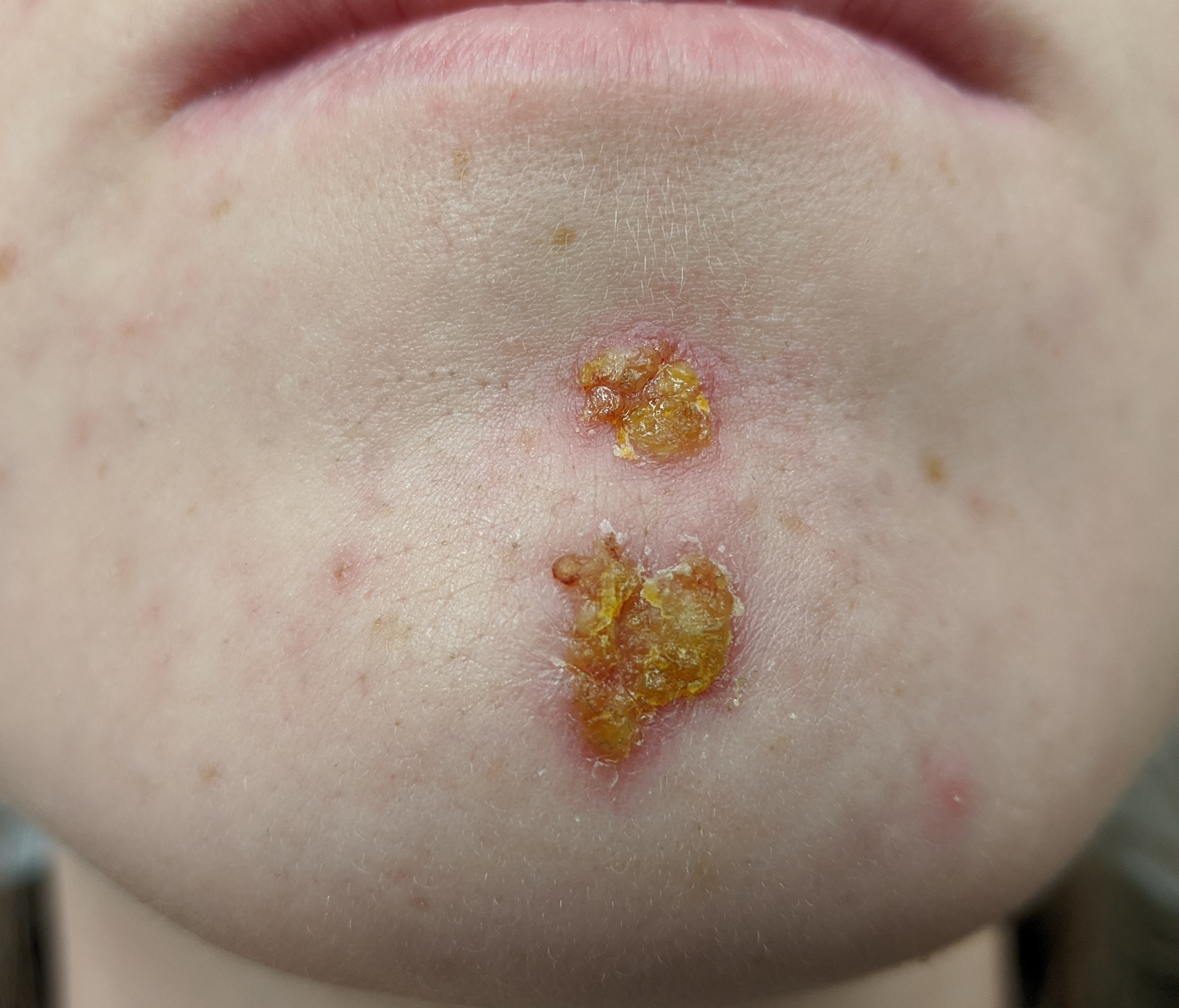
- Impetigo (epidermis): honey-colored crusts (non-bullous, S. aureus > S. pyogenes) or flaccid bullae (bullous, S. aureus exfoliative toxin cleaving desmoglein-1). Strep sequela = post-streptococcal glomerulonephritis, NOT rheumatic fever (skin strep does not cause ARF). Tx: topical mupirocin; oral if extensive.
- Erysipelas (upper dermis/lymphatics): raised, sharply demarcated, fiery-red, fever; S. pyogenes. Tx: penicillin.
- Cellulitis (deep dermis/subcutis): flat, poorly demarcated, warm, tender; S. aureus/S. pyogenes. Purulent or risk factors → cover MRSA (TMP-SMX, doxycycline, or clindamycin).
- Folliculitis / furuncle / carbuncle: S. aureus; hot-tub folliculitis = Pseudomonas aeruginosa.
- SSSS: exfoliative toxin cleaves desmoglein-1 → split at stratum granulosum; Nikolsky +, spares mucosa, infants/young children; blister cultures sterile.
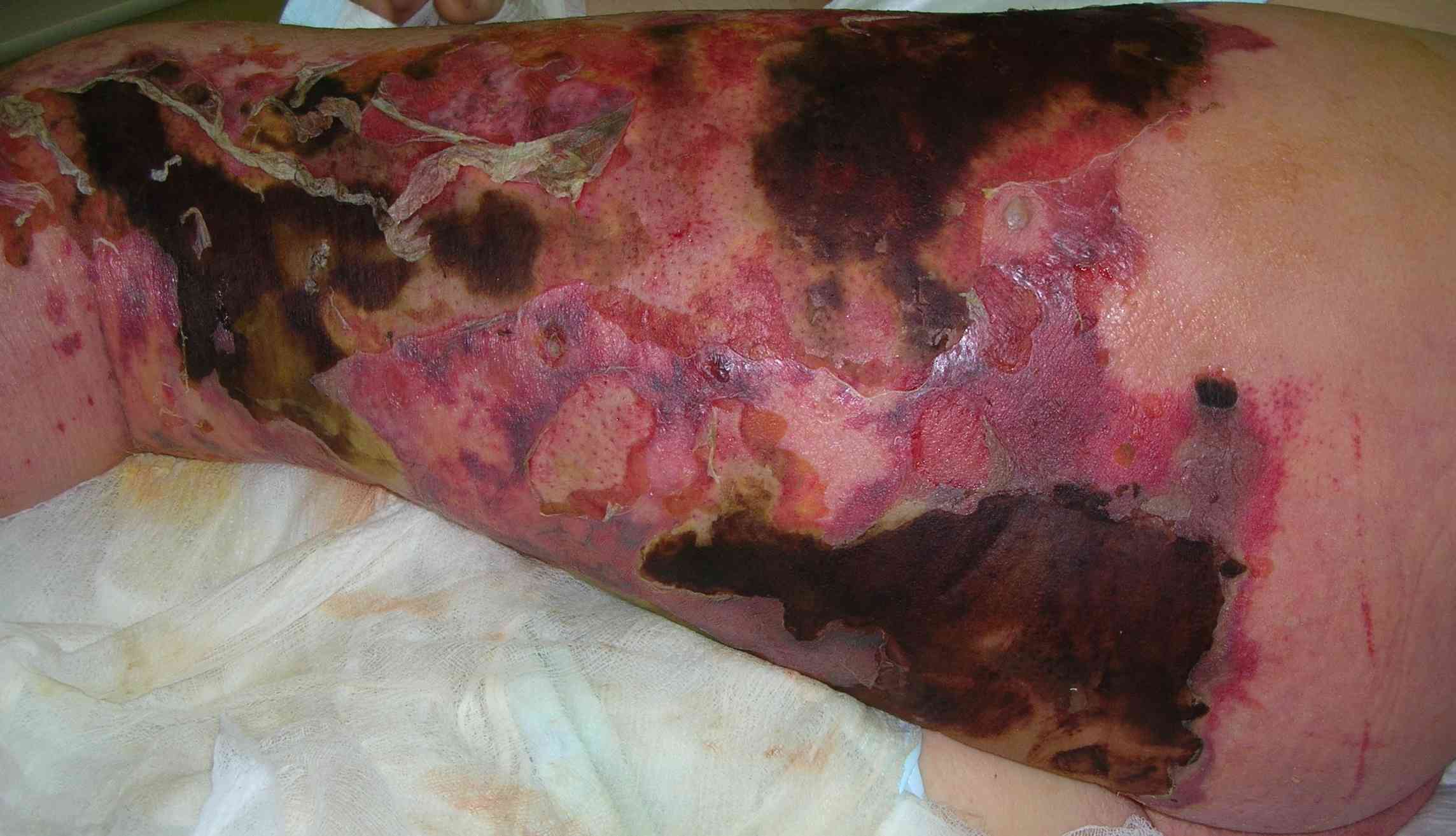
- Necrotizing fasciitis: GAS (type II) or polymicrobial (type I, diabetics); pain out of proportion, crepitus, gray "dishwater" fluid → surgical emergency.
Bacterial Infections by Skin Depth
| Infection | Depth | Key organism | Buzzword | First-line Tx |
|---|---|---|---|---|
| Impetigo | Epidermis | S. aureus (S. pyogenes) | Honey-colored crust | Topical mupirocin |
| Erysipelas | Upper dermis / lymphatics | S. pyogenes | Raised, sharp borders | Penicillin / amoxicillin |
| Cellulitis | Deep dermis / subcutis | S. aureus, S. pyogenes | Flat, ill-defined, warm | Cephalexin (purulent/MRSA → TMP-SMX) |
| Necrotizing fasciitis | Fascia / muscle | GAS or polymicrobial | Pain out of proportion, crepitus | Surgical debridement + broad abx |
Vignette: A 58-year-old man with diabetes has 24 hours of a rapidly spreading, dusky, exquisitely tender leg. Pain is far out of proportion to the modest overlying erythema. He is febrile, tachycardic, and hypotensive; the skin is tense with palpable crepitus, and probing expresses foul, gray "dishwater" fluid. Labs: leukocytosis, high CRP, hyponatremia, rising creatinine and lactate.
Diagnosis: Necrotizing fasciitis (type II GAS, or type I polymicrobial in a diabetic).
Next best step:
- Immediate surgical exploration and debridement — do NOT delay for imaging.
- Empiric vancomycin (or linezolid) + piperacillin-tazobactam + clindamycin — clindamycin (a protein-synthesis inhibitor) suppresses streptococcal exotoxin/M-protein production and stays active against high-inoculum, slowly dividing organisms, overcoming the penicillin "Eagle effect."
- Aggressive fluid resuscitation / ICU support.
CT/MRI (fascial gas, edema) or the LRINEC score can support the diagnosis but must never postpone the OR when suspicion is high.
- HSV-1 / HSV-2: painful grouped vesicles on an erythematous base, recurrent. Tzanck smear → multinucleated giant cells (also VZV); PCR is most sensitive/confirmatory. Latency in sensory ganglia. Variants: herpetic whitlow (finger), gladiatorum (wrestlers).
- Eczema herpeticum (Kaposi varicelliform eruption): HSV superinfecting atopic dermatitis → monomorphic, punched-out erosions, ill child — emergency → acyclovir (IV if severe/periocular).
- VZV — varicella: pruritic vesicles in different stages simultaneously, centripetal; "dew-drop on a rose petal."
- VZV — zoster: dermatomal, respects the midline; reactivation from dorsal-root/cranial ganglia. Hutchinson sign (vesicle on nasal tip → V1/ocular risk), Ramsay Hunt (facial palsy + ear vesicles, geniculate ganglion). Tx: valacyclovir; postherpetic neuralgia risk.
- Molluscum contagiosum (poxvirus): umbilicated dome papules with central plug (molluscum bodies). Widespread/facial in an adult → consider HIV.

Vignette: A 3-year-old with known atopic dermatitis develops, over 2 days, clusters of monomorphic, "punched-out" erosions with hemorrhagic crusts coalescing over the face and eczematous neck; a few show central umbilication. He is febrile and looks unwell. A Tzanck smear from a fresh vesicle base shows multinucleated giant cells.
Diagnosis: Eczema herpeticum (disseminated HSV over atopic dermatitis) = Kaposi varicelliform eruption. Do not mislabel as impetigo.
Next best step:
- Start acyclovir promptly (IV if systemic, extensive, or periocular) — antivirals are the priority, not antibiotics alone.
- Confirm with HSV PCR / DFA.
- Ophthalmology if periocular (herpes keratitis risk).
- Treat any bacterial superinfection.
SSSS vs. Toxic Epidermal Necrolysis (TEN)
| Feature | SSSS | TEN |
|---|---|---|
| Cause | S. aureus exfoliative toxin (protease that cleaves desmoglein-1) | Drug reaction (keratinocyte apoptosis) |
| Typical age | Infants / young children | Adults |
| Cleavage plane | Stratum granulosum (superficial) | Dermo-epidermal junction (full-thickness) |
| Mucosa | Spared | Involved (≥2 sites) |
| Nikolsky sign | Positive | Positive |
| Biopsy | Superficial split, sparse infiltrate | Full-thickness epidermal necrosis |
| Treatment | Anti-staph abx (nafcillin; vancomycin if MRSA) + supportive | Stop drug; burn-unit / supportive care |
Blistering / split-level (real classics):
- Desmoglein-1 = superficial (granular layer), spares mucosa (dsg-3 compensates there) → SSSS toxin and pemphigus foliaceus.
- Desmoglein-3 = deep + mucosa → pemphigus vulgaris. Hook: "3 = deep and in the mouth."
- Nikolsky + (skin shears with light pressure) → SSSS, TEN/SJS, pemphigus vulgaris (bullous pemphigoid is Nikolsky −).
Buzzword → diagnosis:
- Honey-colored crust → impetigo
- Dew-drop on a rose petal → varicella
- Umbilicated papule → molluscum
- Grouped vesicles + Tzanck giant cells → HSV / VZV
- Dermatomal rash respecting the midline → zoster
- Pain out of proportion + crepitus → necrotizing fasciitis (screen with LRINEC)
Practice Dermatology now
Board-style questions, spaced-repetition flashcards, and a Socratic AI tutor — free to start.