Autonomic Nervous System Anatomy
A board-focused tour of autonomic anatomy: the two-neuron sympathetic (thoracolumbar) vs parasympathetic (craniosacral) wiring, the sympathetic chain with its splanchnic and adrenal-medulla pathways, the four cranial parasympathetic ganglia, and the classic lesion vignettes — Horner syndrome, compressive vs ischemic CN III palsy, Adie pupil, and Frey syndrome.
How the autonomic nervous system is wired
The autonomic nervous system (ANS) is the visceral motor system — it drives smooth muscle, cardiac muscle, and glands automatically. It has two divisions with opposing tone: sympathetic (thoracolumbar, "fight-or-flight") and parasympathetic (craniosacral, "rest-and-digest").
The single most testable structural fact: every autonomic efferent is a two-neuron chain. A preganglionic neuron in the CNS synapses in an autonomic ganglion onto a postganglionic neuron that reaches the target — unlike the single lower motor neuron of somatic movement. All preganglionic fibers of both divisions release ACh onto nicotinic (N_N) receptors; the divisions differ at the second synapse. A third division, the enteric nervous system (submucosal Meissner + myenteric Auerbach plexuses), runs the gut semi-autonomously and is modulated by the other two. Boards test the outflow levels, the ganglion locations, the neurotransmitter exceptions, and — above all — the lesions.

- Sympathetic = thoracolumbar: preganglionic cell bodies in the intermediolateral cell column (lateral horn), T1–L2 (some texts L3)
- Parasympathetic = craniosacral: cranial nerves III, VII, IX, X and sacral S2–S4
- All preganglionics (both divisions) → ACh → nicotinic (N_N) on the postganglionic cell
- Sympathetic postganglionic → norepinephrine (α/β) — key exception: sweat glands use ACh → muscarinic (sympathetic cholinergic)
- Parasympathetic postganglionic → ACh → muscarinic
- Adrenal medulla = modified sympathetic ganglion: preganglionic (greater splanchnic) fibers synapse directly on chromaffin cells — no postganglionic neuron — releasing ~80% epinephrine / 20% NE into blood
- Sympathetic: short preganglionic / long postganglionic (ganglia near the cord)
- Parasympathetic: long preganglionic / short postganglionic (ganglia in or on the target organ)
- White ramus communicans = myelinated preganglionic, present only T1–L2; gray ramus communicans = unmyelinated postganglionic, present at every spinal level
- Top clinical pearl: a fixed, dilated ("blown") pupil = compressive CN III (surgical — PComm aneurysm / uncal herniation); a pupil-sparing CN III palsy = ischemic (diabetic microvascular); the triad of partial ptosis + miosis + anhidrosis = Horner
Sympathetic outflow: the three fates and the splanchnic nerves
Preganglionic sympathetic axons leave the cord in the ventral root, join the spinal nerve, then peel off through the white ramus communicans into the paravertebral sympathetic chain (trunk). Once in the chain, a fiber has three options:
- Synapse at the same level in a paravertebral (chain) ganglion.
- Ascend or descend the chain, then synapse — e.g., head/neck sympathetics ascend to the superior cervical ganglion (there are no white rami above T1, so they must travel up).
- Pass straight through without synapsing and exit as a splanchnic nerve to a prevertebral (collateral) ganglion on the aorta.
Postganglionic fibers for the body wall and limbs re-enter the spinal nerve via the gray ramus communicans to reach sweat glands, arrector pili, and vessels. The abdominopelvic splanchnics — greater (T5–T9), lesser (T10–T11), least (T12) — synapse in the celiac, superior mesenteric, aorticorenal, and inferior mesenteric ganglia for the gut. Special case: greater-splanchnic preganglionic fibers pass through the celiac ganglion and synapse directly on the adrenal medulla. Classic trap: sacral splanchnic nerves are sympathetic, whereas pelvic splanchnic nerves (S2–S4) are parasympathetic.
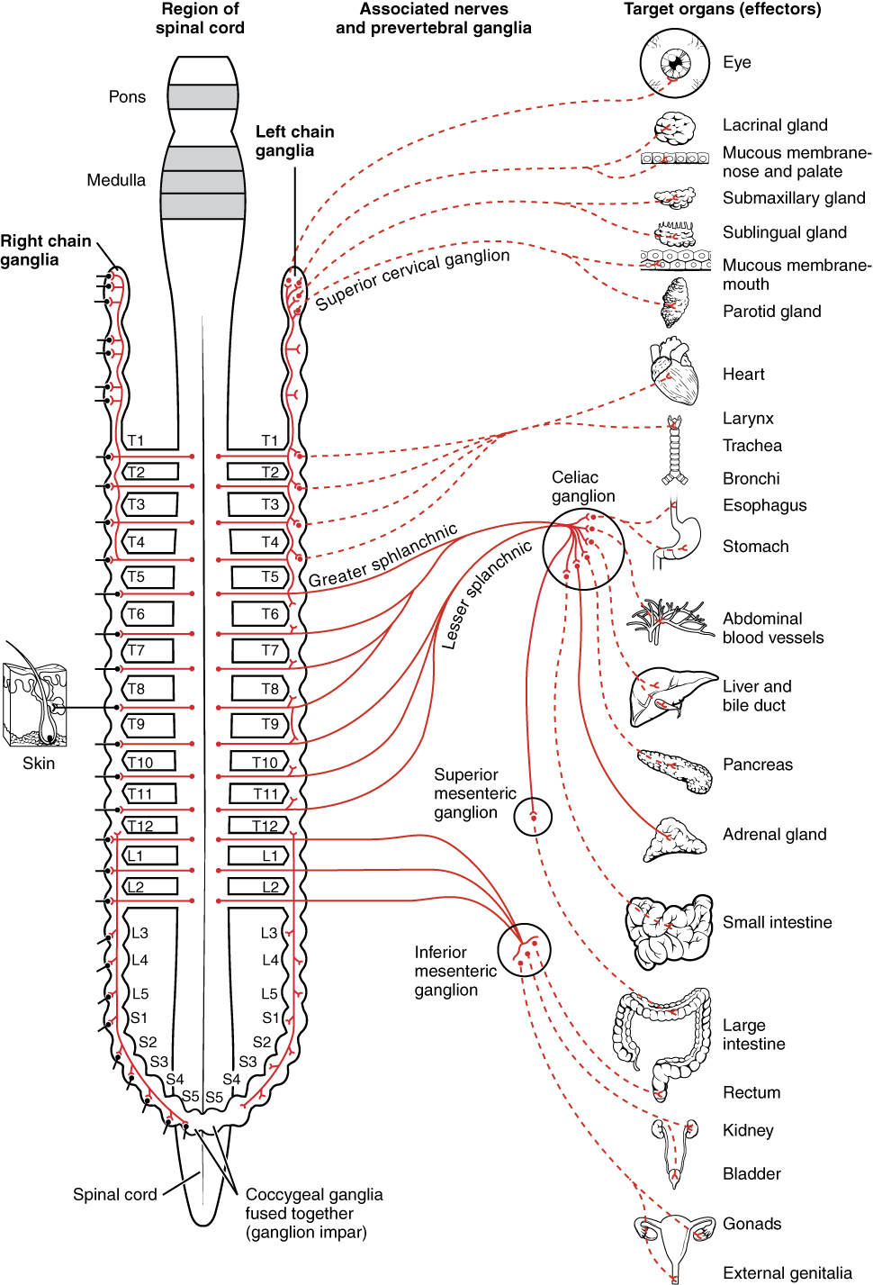
Sympathetic vs parasympathetic at a glance
| Feature | Sympathetic | Parasympathetic |
|---|---|---|
| Outflow | Thoracolumbar T1–L2 | Craniosacral (CN III, VII, IX, X; S2–S4) |
| Preganglionic cell body | Intermediolateral cell column | Brainstem CN nuclei / sacral parasympathetic nucleus (S2–S4) |
| Ganglion location | Near cord (para-/prevertebral) | Near or within the target organ |
| Pre : post fiber length | Short : long | Long : short |
| Postganglionic transmitter | NE (sweat glands = ACh) | ACh |
| Postganglionic receptor | α, β adrenergic | Muscarinic |
| Pupil | Dilates (radial m., mydriasis) | Constricts (sphincter, miosis) |
| Heart rate | ↑ | ↓ |
| Bronchi | Dilate | Constrict + secretions |
| GI motility | ↓ (sphincters contract) | ↑ secretion & peristalsis |
| Bladder | Stores (internal sphincter contracts) | Voids (detrusor contracts) |
| Sexual | Ejaculation | Erection |
The four cranial parasympathetic ganglia
| Ganglion | Preganglionic CN | Route to ganglion | Target (postganglionic) |
|---|---|---|---|
| Ciliary | CN III (Edinger–Westphal n.) | Inferior division of III → short ciliary nn. | Sphincter pupillae (miosis) + ciliary muscle (accommodation) |
| Pterygopalatine | CN VII | Greater petrosal → n. of pterygoid canal | Lacrimal gland + mucous glands of nose/palate |
| Submandibular | CN VII | Chorda tympani → lingual n. | Submandibular + sublingual salivary glands |
| Otic | CN IX | Lesser petrosal → auriculotemporal n. | Parotid gland |
- Cranial parasympathetic outflow = 3, 7, 9, 10.
- Muscarinic (cholinergic) excess — organophosphate / nerve-agent toxidrome — "DUMBELS": Diarrhea, Urination, Miosis, Bradycardia / Bronchospasm / Bronchorrhea (the "killer B's"), Emesis, Lacrimation, Salivation/Sweating. (Overlapping short form SLUDGE: Salivation, Lacrimation, Urination, Defecation, GI cramping, Emesis.)
- Horner triad — "PAM": Ptosis, Anhidrosis, Miosis (± apparent enophthalmos).
- Sexual autonomics — "Point and Shoot": Parasympathetic = Point (erection); Sympathetic = Shoot (ejaculation).
- Ganglion → gland: the Otic ganglion (the odd one, from CN IX) serves the parotid; the Submandibular ganglion serves the submandibular/sublingual glands.
Vignette: A smoker with an apical lung mass develops a drooping right upper lid, a constricted right pupil, and a dry, non-sweating right side of the face.
The sympathetic supply to the eye is a three-neuron chain; where it breaks localizes the lesion:
- First-order (central): hypothalamus → descends the brainstem/cord to the ciliospinal center of Budge (C8–T2). Lesion: lateral medullary (Wallenberg) stroke, syringomyelia.
- Second-order (preganglionic): C8–T2 → arches over the lung apex → superior cervical ganglion. Lesion: Pancoast (apical lung) tumor, cervical rib.
- Third-order (postganglionic): superior cervical ganglion → travels along the internal carotid artery → orbit. Lesion: internal carotid dissection, cavernous sinus lesion.
Deficit: ptosis (paralysis of the sympathetic superior tarsal / Müller muscle — partial, unlike the complete ptosis of a CN III palsy), miosis (unopposed sphincter pupillae; the pupil dilates poorly, so anisocoria is worse in dim light), and anhidrosis of the ipsilateral face. Sweating is spared in third-order (postganglionic) lesions distal to the carotid bifurcation, because facial sudomotor fibers follow the external carotid.

Key anatomy: the parasympathetic (pupilloconstrictor) fibers of CN III travel on the outer surface of the nerve, while the somatic motor fibers to the extraocular muscles run in the core. That layering explains two opposite vignettes:
- Compressive CN III palsy (posterior communicating artery aneurysm, uncal/transtentorial herniation): the superficial parasympathetic fibers are squeezed first → a fixed, dilated "blown" pupil, with the eye "down and out" and ptosis. A blown pupil flags a surgical/compressive third-nerve palsy.
- Ischemic CN III palsy (diabetes, hypertension — vasa nervorum infarct): the central somatic fibers infarct but the pupil is spared ("pupil-sparing"), because the peripheral parasympathetic fibers keep their blood supply.
- Adie (tonic) pupil: damage to the ciliary ganglion / short ciliary nerves → a large pupil, poorly reactive to light with a slow "tonic" near response (light-near dissociation); benign, classically young women.
- Frey syndrome: after parotid surgery, regenerating parasympathetic fibers (auriculotemporal nerve, from the otic ganglion) misdirect onto facial sweat glands → gustatory sweating/flushing over the cheek while eating.
Practice Anatomy now
Board-style questions, spaced-repetition flashcards, and a Socratic AI tutor — free to start.