Asthma & COPD
A boards-focused Asthma vs COPD lesson running pathophysiology → presentation buzzwords → diagnosis (PFTs/imaging/ABG) → next-best-step management for STEP 1 and STEP 2 CK. Emphasizes reversibility testing, the falsely-normal PaCO2 trap, and GINA/GOLD-current therapy.
Two obstructive diseases, one testable distinction
Asthma and COPD are the two great obstructive diseases (both show ↓ FEV1/FVC), but boards hinge on reversibility and who gets it.
Asthma = chronic Th2 / eosinophilic airway inflammation → reversible bronchospasm and airway hyperresponsiveness, driven by mast cells, eosinophils, and IgE. Triggers: allergens, exercise, cold air, viral URIs, and aspirin/NSAIDs (AERD / Samter triad = asthma + nasal polyps + aspirin sensitivity).
COPD = smoking-driven, largely irreversible airflow limitation from chronic bronchitis (mucous-gland hyperplasia, ↑ Reid index) plus emphysema (alveolar-wall destruction, loss of elastic recoil). Driven by neutrophils, macrophages, and CD8+ T cells. A young nonsmoker with lower-lobe panacinar emphysema ± early liver disease → suspect alpha-1 antitrypsin (A1AT) deficiency.
- Asthma sputum: Curschmann spirals (mucus casts), Charcot-Leyden crystals (eosinophil galectin-10)
- AERD: aspirin/NSAIDs block COX → shunt arachidonate to leukotrienes → bronchospasm; manage with leukotriene modifiers ± aspirin desensitization
- Emphysema types: centriacinar (smoking, upper lobes) vs panacinar (A1AT, lower lobes)
- Chronic bronchitis = productive cough ≥ 3 months/yr × 2 consecutive years
- Asthma presentation: episodic wheeze/cough worse at night/early morning; often normal between attacks
- COPD presentation: progressive dyspnea, barrel chest, prolonged expiration, pursed-lip breathing; advanced disease → cor pulmonale (JVD, hepatomegaly, peripheral edema)
The classic COPD phenotypes — match the letters:
- Pink Puffer = emPhysema → Pursed-lip breathing, Pink (keeps SpO2 up by hyperventilating), thin/cachectic, barrel chest, ↓ DLCO, quiet chest
- Blue Bloater = chronic Bronchitis → Blue (cyanotic/hypoxemic), Bloated (edema from cor pulmonale), productive cough, CO2 retention
Hook: the P's track with emPhysema; the B's track with Bronchitis. Real patients overlap, but exam vignettes lean on these caricatures.
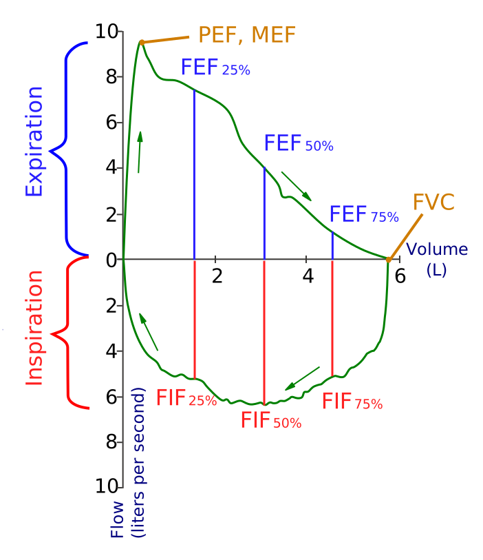
Confirm obstruction with spirometry, then test reversibility.
- Both: post-bronchodilator FEV1/FVC < 0.70
- Asthma reversibility: FEV1 improves ≥ 12% AND ≥ 200 mL after bronchodilator. Normal spirometry but classic history? → methacholine challenge (≥ 20% ↓ FEV1 = hyperresponsive)
- Lung volumes (air trapping/hyperinflation): ↑ TLC, RV, FRC
- DLCO: ↓ in emphysema; normal or ↑ in asthma — a key discriminator
- CXR: emphysema → hyperinflation, flattened diaphragms, ↑ retrosternal airspace, small vertical heart, bullae; asthma CXR often normal
- Labs: peripheral eosinophilia + ↑ IgE (allergic asthma); order A1AT level in early-onset or nonsmoker COPD
- ABG in severe asthma attack: early respiratory alkalosis; a normalizing or rising PaCO2 = fatigue → impending respiratory failure
Asthma vs COPD at a glance
| Feature | Asthma | COPD |
|---|---|---|
| Typical onset | Childhood / young adult | > 40 yr, smoker |
| Reversibility | Reversible (≥ 12% & ≥ 200 mL ↑ FEV1) | Largely irreversible |
| FEV1/FVC | < 0.70 in attack, normal between | < 0.70 fixed (post-BD) |
| DLCO | Normal or ↑ | ↓ (emphysema) |
| Inflammatory cells | Eosinophils, Th2, IgE | Neutrophils, CD8+, macrophages |
| Challenge test | Methacholine → ↓ FEV1 | Not used |
| First-line controller | ICS (± formoterol) | LAMA ± LABA |

Vignette: A 22-year-old with asthma has 6 hours of worsening dyspnea. RR 32, accessory-muscle use, speaking in single words, peak flow 40% predicted, SpO2 90%. Initial ABG PaCO2 was 26; a repeat now shows PaCO2 41 ("normal") and he appears exhausted.
- Dx: severe / near-fatal asthma exacerbation
- Trap: a normal PaCO2 in a tachypneic asthmatic is falsely reassuring — it means he can no longer sustain hyperventilation → impending respiratory failure
- Next best step: prepare for endotracheal intubation (this near-arrest picture is where NIPPV is unreliable). Concurrently give O2, continuous nebulized SABA (albuterol) + ipratropium, systemic corticosteroids, and IV magnesium sulfate for severe/refractory cases
Vignette: A 68-year-old with a 50-pack-year history has 3 days of increased dyspnea and purulent sputum. RR 28, drowsy. ABG on 2 L O2: pH 7.28, PaCO2 68, PaO2 55.
- Dx: acute COPD exacerbation with acute (on chronic) hypercapnic respiratory failure (respiratory acidosis)
- Next best step: noninvasive positive-pressure ventilation (BiPAP) — in COPD with respiratory acidosis it reduces intubation rate and mortality (trial it with close monitoring even when hypercapnic drowsiness is present)
- Also: controlled O2 targeting SpO2 88–92% (over-oxygenation worsens CO2 retention via V/Q mismatch + Haldane effect), nebulized SABA + ipratropium, systemic corticosteroids (~5-day course), and antibiotics when ≥ 2 of ↑ dyspnea / ↑ sputum volume / ↑ sputum purulence (Anthonisen criteria)
Asthma (GINA):
- Controller cornerstone = inhaled corticosteroid (ICS); GINA's preferred reliever is now low-dose ICS-formoterol, NOT SABA alone
- Step up: add LABA → ↑ ICS dose → add LAMA / LTRA → biologics for severe disease (anti-IgE omalizumab; anti-IL-5 mepolizumab/reslizumab, anti-IL-5Rα benralizumab; anti-IL-4Rα dupilumab, blocking IL-4 and IL-13)
COPD (GOLD):
- Mainstay = long-acting bronchodilators LAMA ± LABA; add ICS if blood eosinophils ≥ 300 or asthma overlap (ICS raises pneumonia risk)
- Proven to ↓ mortality: smoking cessation and long-term O2 therapy in chronic hypoxemia (PaO2 ≤ 55 mmHg / SpO2 ≤ 88%, or ≤ 59 with cor pulmonale/polycythemia); lung volume reduction surgery helps a selected upper-lobe-predominant subgroup
- Vaccinate: influenza, pneumococcal, COVID-19, plus RSV/pertussis per age
Practice Pulmonary now
Board-style questions, spaced-repetition flashcards, and a Socratic AI tutor — free to start.