ARDS & Acute Respiratory Failure
A Step 2 CK-focused pulmonology lesson on ARDS and acute respiratory failure, moving from pathophysiology (diffuse alveolar damage, shunt physiology) through the Berlin diagnostic criteria and cardiogenic-edema differential to evidence-based ventilator management. Emphasizes classic vignette buzzwords and next-best-step decisions (intubation with lung-protective ventilation, BiPAP for hypercapnic failure, prone positioning for refractory hypoxemia).
Overview & Pathophysiology
Acute respiratory failure is inadequate gas exchange, defined on ABG as PaO₂ < 60 mmHg (Type 1, hypoxemic) and/or PaCO₂ > 50 mmHg with acidemia (Type 2, hypercapnic). Type 1 arises from V/Q mismatch or shunt (pneumonia, edema, PE, ARDS); Type 2 from alveolar hypoventilation (COPD, asthma, neuromuscular disease, opioid overdose).
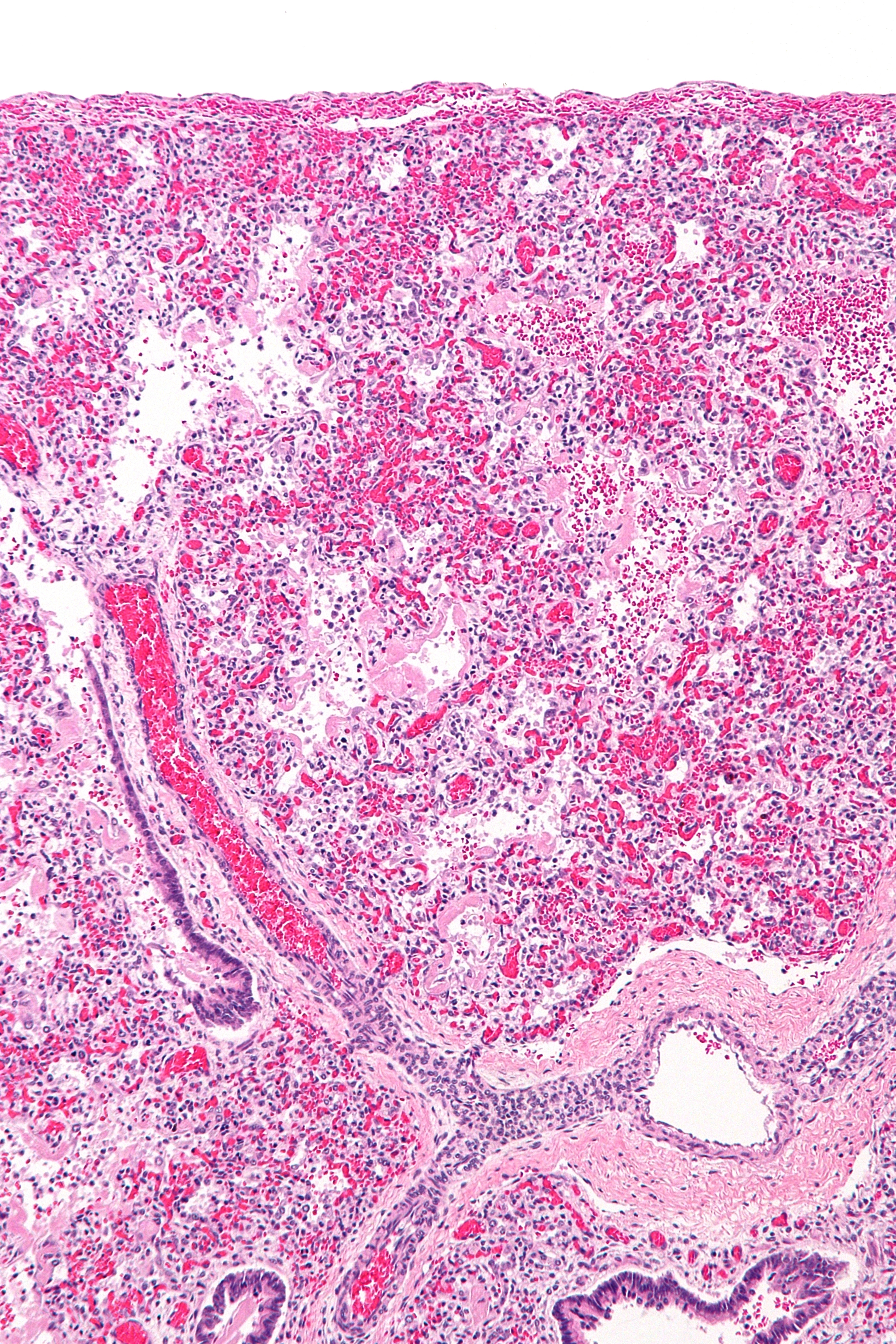
ARDS is the severe end of hypoxemic failure — an acute, diffuse inflammatory lung injury causing non-cardiogenic pulmonary edema. A systemic or pulmonary insult activates neutrophils and cytokines that injure the alveolar–capillary membrane. Increased permeability floods alveoli with protein-rich exudate, inactivates surfactant, and forms hyaline membranes — the histologic hallmark diffuse alveolar damage (DAD). Lungs become stiff (low compliance), and blood passing unventilated flooded alveoli creates intrapulmonary shunt → hypoxemia that does not correct with supplemental O₂. It evolves through exudative (days 0–7) → proliferative → fibrotic phases. Common triggers: sepsis (most common), aspiration, pneumonia, pancreatitis, trauma/near-drowning, and multiple transfusions (TRALI).
- Berlin definition — all 4 required:
- Timing: within 1 week of a known insult or new/worsening symptoms
- Imaging: bilateral opacities not fully explained by effusion, collapse, or nodules
- Origin: edema not fully explained by cardiac failure/fluid overload (get echo ± BNP if no clear risk factor)
- Oxygenation (on PEEP or CPAP ≥ 5): by PaO₂/FiO₂ ratio — Mild 200–300, Moderate 100–200, Severe ≤ 100
- ABG: hypoxemia with a widened A–a gradient; early respiratory alkalosis from tachypnea — a rising PaCO₂ is a late, ominous sign of fatigue
- Refractory hypoxemia (little improvement with 100% O₂) reflects shunt — a key ARDS clue
- PA catheter/PCWP is no longer required; if measured, PCWP ≤ 18 favors ARDS over cardiogenic edema
- Normal heart size and absent cephalization/Kerley lines on CXR argue against a cardiac cause
Type 1 vs Type 2 Respiratory Failure
| Feature | Type 1 (Hypoxemic) | Type 2 (Hypercapnic) |
|---|---|---|
| ABG | PaO₂ < 60, PaCO₂ normal/low | PaO₂ < 60, PaCO₂ > 50 + acidemia |
| Mechanism | V/Q mismatch, shunt | Alveolar hypoventilation |
| Classic causes | ARDS, pneumonia, pulmonary edema, PE | COPD/asthma, opioid OD, neuromuscular (GBS, MG), obesity-hypoventilation |
| A–a gradient | Increased | Normal (pure hypoventilation) or increased |
| First-line support | O₂ ± PEEP; treat cause | NIV (BiPAP); reverse cause |
ARDS vs Cardiogenic Pulmonary Edema
| Feature | ARDS (non-cardiogenic) | Cardiogenic edema |
|---|---|---|
| Mechanism | ↑ capillary permeability | ↑ hydrostatic pressure |
| PCWP | ≤ 18 (normal) | > 18 (elevated) |
| Heart size | Normal | Enlarged |
| CXR extras | Peripheral patchy opacities; no cephalization | Cephalization, Kerley B lines, effusions |
| BNP | Low/normal | Elevated (> 500) |
| Edema fluid | Protein-rich exudate | Protein-poor transudate |
| Diuresis response | Minimal | Improves |
Vignette: A 58-year-old man admitted for severe gallstone pancreatitis becomes acutely dyspneic on hospital day 2. RR 34, SpO₂ 85% on a non-rebreather, diffuse crackles. CXR: bilateral alveolar infiltrates. Bedside echo: normal EF, no volume overload. ABG on FiO₂ 1.0: PaO₂ 100 → PaO₂/FiO₂ = 100.
Diagnosis: Severe ARDS (Berlin — onset < 1 wk, bilateral opacities, non-cardiogenic, P/F ≤ 100). Pancreatitis is the systemic trigger; the O₂-refractory hypoxemia reflects shunt.
Next best step: Intubate and start lung-protective ventilation — low tidal volume 6 mL/kg predicted body weight, plateau pressure < 30 cmH₂O, titrate PEEP, permissive hypercapnia (keep pH > 7.2) — plus conservative fluids and treatment of the pancreatitis.
If still refractory (P/F < 150): the next step is prone positioning (mortality benefit), then neuromuscular blockade, with VV-ECMO as rescue.

Vignette: A 67-year-old with COPD has 3 days of worsening cough and dyspnea. He uses accessory muscles and is drowsy but rousable. ABG: pH 7.28, PaCO₂ 68, PaO₂ 56, HCO₃⁻ 30.
Diagnosis: Acute-on-chronic Type 2 (hypercapnic) respiratory failure from a COPD exacerbation — acute respiratory acidosis superimposed on a chronically compensated baseline.
Next best step: Noninvasive positive-pressure ventilation (BiPAP) — first-line, it reduces intubation and mortality — plus bronchodilators, systemic steroids, and controlled O₂ targeting SpO₂ 88–92% to avoid worsening CO₂ retention.
Intubate instead if the patient is comatose / cannot protect the airway, is apneic or hemodynamically unstable, or fails a NIV trial. Avoid over-oxygenation, which worsens hypercapnia via V/Q mismatch (loss of hypoxic vasoconstriction) and the Haldane effect.
- Treat the underlying cause (e.g., antibiotics/source control for sepsis) — the only therapy that truly reverses ARDS
- Lung-protective ventilation (ARDSNet/ARMA): tidal volume 6 mL/kg predicted body weight, plateau pressure < 30 cmH₂O — proven mortality benefit
- Permissive hypercapnia: tolerate a high PaCO₂ as long as pH > 7.2
- PEEP to recruit alveoli and reduce shunt; target SpO₂ 88–95% / PaO₂ 55–80
- Conservative fluid strategy (FACTT) → more ventilator-free days
- Severe ARDS (P/F < 150): prone positioning (PROSEVA — mortality benefit); consider early neuromuscular blockade (ACURASYS suggested benefit; ROSE did not — hence selective use)
- VV-ECMO for hypoxemia refractory to the above
- Dexamethasone may reduce ventilator days/mortality (especially COVID-19 ARDS); routine steroids otherwise remain debated
- Avoid high tidal volumes (barotrauma/VILI) and liberal fluids
Classic framework — pair each cause with its A–a gradient:
- Low inspired O₂ (high altitude) — A–a normal
- Hypoventilation (opioids, COPD) — A–a normal
- V/Q mismatch (PE, pneumonia) — A–a ↑, corrects with O₂
- Diffusion limitation (ILD, emphysema) — A–a ↑, corrects with O₂
- Right-to-left shunt (ARDS, atelectasis, intracardiac) — A–a ↑, does NOT correct with O₂
Board pearl: Only a shunt fails to correct with 100% O₂ — this is why ARDS causes refractory hypoxemia. A normal A–a gradient points to hypoventilation or low FiO₂; a widened A–a gradient localizes the problem to the lung (V/Q, diffusion, or shunt).
Practice Pulmonary now
Board-style questions, spaced-repetition flashcards, and a Socratic AI tutor — free to start.