Approach to Common Lab Abnormalities
A next-best-step approach to the electrolyte and acid-base abnormalities Step 2 CK loves — hyponatremia (osmolality → volume status), hyperkalemia (ECG → calcium first), high anion-gap acidosis (MUDPILES), and hypercalcemia (PTH-driven) — pairing each decisive confirmatory lab with the first correct action.
The Approach: Confirm → Classify → Act
Lab abnormalities are the board's favorite launch point for a next-best-step question. The winning move is almost never to treat the number reflexively — it is to (1) confirm it's real (repeat the value; exclude artifact like pseudohyperkalemia from hemolysis, or normal-osmolality pseudohyponatremia from severe hyperlipidemia/paraproteins), (2) classify with a small set of confirmatory labs, and (3) act on the classification.
Each abnormality has one decisive branch point:
- Sodium → serum osmolality, then volume status.
- Potassium → an ECG before anything else.
- Metabolic acidosis → the anion gap.
- Calcium → the PTH.
Master these branch points and most electrolyte/acid-base vignettes collapse into a single correct action.
- Hyponatremia: check serum osmolality first → if low (true hypotonic), assess volume status, then urine osm and urine Na.
- Correction limit: in chronic hyponatremia, raise serum Na by ≤8 mEq/L per 24 h (goal ~4–6) to avoid osmotic demyelination syndrome (central pontine myelinolysis).
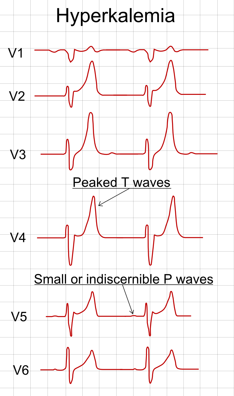
- Hyperkalemia: get an ECG immediately; if ECG changes → IV calcium gluconate to stabilize the myocardium (it does not lower K⁺).
- Metabolic acidosis: anion gap = Na − (Cl + HCO₃); high gap → MUDPILES. Check respiratory compensation with Winter's formula (expected pCO₂ = 1.5×HCO₃ + 8 ± 2).
- Hypercalcemia: the deciding lab is PTH — high/inappropriately normal = primary hyperparathyroidism; low/suppressed = malignancy or vitamin D excess.
- Always stop the offending drug: thiazides/other diuretics (Na⁺); ACEi/ARB, NSAIDs, K⁺-sparing diuretics (K⁺); thiazides, lithium (Ca²⁺).
Hypotonic Hyponatremia by Volume Status
| Volume status | Urine Na | Classic causes | First move |
|---|---|---|---|
| Hypovolemic | <20 (extrarenal); >20 if renal | Vomiting/diarrhea, diuretics, primary adrenal insufficiency | Isotonic (0.9%) saline |
| Euvolemic | >30 | SIADH, hypothyroidism, glucocorticoid deficiency, psychogenic polydipsia (dilute urine) | Fluid restriction |
| Hypervolemic | <20 | Heart failure, cirrhosis, nephrotic syndrome | Water + salt restriction ± loop diuretic |
Vignette: A 64-year-old smoker with a hilar mass has Na 118 mEq/L, is clinically euvolemic, serum osm 250 mOsm/kg (low), urine osm 480, urine Na 55; TSH and morning cortisol are normal.
Diagnosis: SIADH — here paraneoplastic from small cell lung cancer. Inappropriately concentrated urine (osm >100) plus elevated urine Na in a euvolemic, hypotonic patient (thyroid/adrenal excluded) is the classic pattern.
Next step (asymptomatic/mild): fluid restriction. Severe symptoms (seizures, obtundation): hypertonic (3%) saline, correcting ≤8 mEq/L in 24 h. Chronic/refractory: salt tablets, loop diuretic, or a vaptan (ADH-receptor antagonist). Also treat the underlying malignancy. Other SIADH triggers to recognize: CNS insults, pneumonia, and drugs (SSRIs, carbamazepine).
- First exclude pseudohyperkalemia (hemolyzed sample, prolonged tourniquet/fist-clenching, marked thrombocytosis/leukocytosis) — repeat the draw if the patient is well with no cause.
- ECG progression: peaked T waves → flattened P → widened QRS → sine wave → arrest.
- Management order:
- IV calcium gluconate — membrane stabilization, immediate, when ECG changes are present; does NOT lower K⁺.
- Shift K⁺ intracellularly: insulin + glucose and albuterol (add bicarbonate only if acidemic).
- Remove K⁺ from body: loop diuretic, GI binder (patiromer or sodium polystyrene sulfonate), and hemodialysis — definitive, especially in renal failure.
- Stop the culprits: ACEi/ARB, K⁺-sparing diuretics (spironolactone), NSAIDs, trimethoprim, and potassium supplements.
Vignette: A hemodialysis patient who missed two sessions presents with weakness. K⁺ is 7.4 mEq/L and the ECG shows peaked T waves with a widening QRS.
Next best step: IV calcium gluconate NOW to stabilize the cardiac membrane — before insulin/glucose or dialysis. It acts within minutes and buys time, but it does not lower the serum potassium.
Then: insulin + glucose (± albuterol) to drive K⁺ into cells, and arrange urgent hemodialysis as the definitive removal in a patient with ESRD (shifting agents only redistribute; the total-body burden is unchanged).
Classic distractor: choosing insulin/glucose first when ECG changes are present. When the ECG is abnormal, membrane protection comes first.
MUDPILES — causes of a high anion-gap metabolic acidosis:
- M — Methanol
- U — Uremia
- D — Diabetic (and alcoholic/starvation) ketoacidosis
- P — Propylene glycol / Paraldehyde
- I — Isoniazid / Iron
- L — Lactic acidosis
- E — Ethylene glycol
- S — Salicylates
Normal-gap (hyperchloremic) acidosis → think diarrhea and renal tubular acidosis (mnemonic HARDASS).
Board trap: salicylate poisoning classically causes a mixed picture — high anion-gap metabolic acidosis plus a primary respiratory alkalosis (tinnitus, fever, tachypnea). A high gap should also prompt checking the osmolar gap (elevated with toxic alcohols: methanol, ethylene glycol).
Hypercalcemia: PTH Decides
| Feature | Primary hyperparathyroidism | Malignancy |
|---|---|---|
| PTH | High or inappropriately normal | Low / suppressed |
| Typical setting | Outpatient, often asymptomatic, chronic | Inpatient, symptomatic, rapid onset |
| Mechanism | Parathyroid adenoma | PTHrP (squamous cell), osteolytic mets, 1,25-OH₂ vitamin D (lymphoma/granuloma) |
| Phosphate | Low | Variable |
Practice Clinical Diagnosis now
Board-style questions, spaced-repetition flashcards, and a Socratic AI tutor — free to start.