Aortic Arch Derivatives
The pharyngeal (aortic) arch arteries — pairs 1, 2, 3, 4, and 6 (the 5th leaves no derivative) — remodel asymmetrically during weeks 4–8 into the definitive great vessels. Their failures generate the board's classic malformations: PDA (left 6th arch), coarctation of the aortic isthmus, interrupted arch (DiGeorge/22q11), and the vascular rings.
The arch arteries and how they remodel
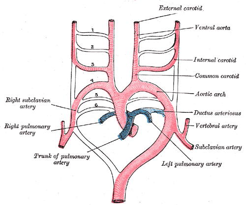
The pharyngeal (aortic) arch arteries are paired vessels running through the pharyngeal arches, connecting the aortic sac (the distal outflow of the truncus arteriosus) to the paired dorsal aortae. Five pairs form and are numbered 1, 2, 3, 4, and 6 — the 5th pair never fully forms and leaves no derivative.
A point the boards love: the arches do not all exist at once. They appear and are remodeled in craniocaudal sequence during weeks 4–8, so by the time the caudal arches mature, the cranial ones have already regressed or transformed. Remodeling is asymmetric — the definitive great vessels come largely from the proximal portions of the right and left arches, while much of the distal right arch and right dorsal aorta involute. That asymmetry is exactly what generates the classic malformations and the odd course of the recurrent laryngeal nerves.
- Five pairs form (1, 2, 3, 4, 6); the 5th regresses / never fully forms → no derivative.
- Arches remodel in craniocaudal sequence, weeks 4–8 — they never all coexist.
- 1st → maxillary artery (branch of external carotid); mostly regresses.
- 2nd → stapedial + hyoid arteries; mostly regresses.
- 3rd = Carotid: common carotid + proximal internal carotid.
- 4th = systemic: LEFT → arch of aorta; RIGHT → proximal right subclavian.
- 6th = pulmonary: proximal pulmonary arteries bilaterally; the LEFT 6th also becomes the ductus arteriosus (→ ligamentum arteriosum after birth).
- Adult vessels arise mostly from the proximal arch segments; distal right arch + right dorsal aorta largely regress — the root cause of left–right asymmetry.
Structure → derivative
| Aortic arch | Adult derivative(s) |
|---|---|
| 1st | Part of the maxillary artery; largely regresses |
| 2nd | Stapedial and hyoid arteries; largely regresses |
| 3rd | Common carotid + proximal internal carotid |
| 4th — LEFT | Arch of the aorta (segment between left common carotid and left subclavian) |
| 4th — RIGHT | Proximal right subclavian artery |
| 5th | No derivative (never fully forms / regresses) |
| 6th — both | Proximal pulmonary arteries |
| 6th — LEFT only | Ductus arteriosus → ligamentum arteriosum |
- 1st arch is MAXimal → maxillary artery.
- Second = Stapedial (2 → Stapedial + hyoid).
- C is the 3rd letter of the alphabet → 3rd arch = Common Carotid + proximal internal Carotid.
- 4th arch = 4 limbs = systemic → arch of aorta (left) + subclavian (right).
- 6th arch = pulmonary + the pulmonary-to-systemic shunt (ductus arteriosus).
Counting trick: run 1, 2, 3, 4, 6 and consciously skip the 5th — that omission is itself a favorite testing point.
Why the recurrent laryngeal nerves are asymmetric
Both recurrent laryngeal nerves (RLNs) begin hooked under the 6th arch arteries. Then asymmetry takes over:
- On the RIGHT, the distal 6th arch (and the 5th) regress, so the right RLN slips upward to loop under the next persisting vessel — the right subclavian artery (4th arch derivative).
- On the LEFT, the 6th arch persists as the ductus / ligamentum arteriosum, so the left RLN stays hooked around the aortic arch at the ligamentum arteriosum.
Board correlate: an aortic arch aneurysm, an enlarged left atrium (classically mitral stenosis), or traction at the ligamentum arteriosum can stretch the left RLN → hoarseness (cardiovocal / Ortner syndrome). The left RLN is likewise at risk with a left-sided PDA.

- PDA ↔ congenital rubella, prematurity, high altitude; continuous machine-like murmur.
- Coarctation ↔ Turner syndrome, bicuspid aortic valve, rib notching, berry aneurysm.
- Interrupted aortic arch (type B) ↔ 22q11.2 deletion / DiGeorge (also truncus arteriosus, tetralogy).
- Double aortic arch / right aortic arch ↔ vascular ring → infant stridor + dysphagia.
- Aberrant right subclavian ↔ dysphagia lusoria (vessel courses behind the esophagus).
- Duct-dependent lesions: keep the duct open with prostaglandin E1 (alprostadil); close a PDA with indomethacin (prostaglandin-synthesis inhibitor).
A 14-year-old girl with short stature, a webbed neck, and primary amenorrhea has BP 155/95 in both arms but 105/60 in the legs, with weak, delayed femoral pulses (radio-femoral delay). Chest X-ray shows notching of the inferior rib margins.
Diagnosis: Coarctation of the aorta — a narrowing of the aortic isthmus, classically juxtaductal (near the ligamentum arteriosum). The upper-vs-lower BP gradient and weak femoral pulses are the signature. Rib notching comes from collateral flow through dilated intercostal arteries eroding the ribs. Classic associations: Turner syndrome and bicuspid aortic valve.
A 3-month-old born at 30 weeks; the mother had a febrile rash with arthralgias and postauricular/suboccipital lymphadenopathy in the first trimester. Exam reveals a continuous, 'machine-like' murmur loudest at the left infraclavicular region, bounding pulses, and a wide pulse pressure.
Diagnosis: Patent ductus arteriosus — the left 6th-arch-derived ductus arteriosus failed to close into the ligamentum arteriosum, leaving a left-to-right shunt. Congenital rubella and prematurity are the classic setups. Indomethacin (inhibits prostaglandin synthesis) promotes closure; prostaglandin E1 keeps the duct open when systemic or pulmonary flow is duct-dependent.
Defect → embryologic mechanism
| Malformation | Embryologic basis | Presentation / association |
|---|---|---|
| Patent ductus arteriosus | Left 6th arch ductus arteriosus fails to close | Continuous machine-like murmur, wide pulse pressure; rubella, prematurity |
| Coarctation of the aorta | Narrowing of the aortic arch/isthmus, juxtaductal | Upper-limb HTN, weak femoral pulses, rib notching; Turner, bicuspid valve |
| Interrupted aortic arch (type B) | Left 4th arch segment (between L common carotid & L subclavian) fails to form | Duct-dependent systemic flow; 22q11.2 / DiGeorge |
| Double aortic arch | Right dorsal aorta fails to regress → both arches persist | Complete vascular ring → infant stridor, dysphagia |
| Aberrant right subclavian (arteria lusoria) | Right 4th arch + proximal right dorsal aorta regress; the persistent distal right dorsal aorta forms the subclavian, which arises as the last branch of the arch behind the esophagus | Dysphagia lusoria |
Practice Embryology now
Board-style questions, spaced-repetition flashcards, and a Socratic AI tutor — free to start.