Acute Leukemias (ALL & AML)
A boards-focused walkthrough of acute leukemias: the shared marrow-failure presentation, then ALL vs AML separated by smear, flow markers, and cytogenetics, anchored to classic vignettes (APL with DIC, pediatric ALL) and next-best-step decisions.
What acute leukemia is
Acute leukemias are clonal malignancies of hematopoietic precursors (blasts) with a block in maturation. Blasts pile up in the marrow and spill into blood, crowding out normal hematopoiesis. Diagnosis classically requires ≥20% blasts in blood or marrow (WHO) — though certain AML-defining translocations [t(15;17), t(8;21), inv(16)] are diagnostic of AML regardless of the blast count.
The shared clinical picture follows directly from marrow failure (pancytopenia):
- Anemia → fatigue, pallor, dyspnea
- Thrombocytopenia → petechiae, mucosal bleeding, easy bruising
- Neutropenia → fever/infection (functionally neutropenic even when total WBC is high)
Onset is acute (days–weeks). Blast infiltration adds bone pain, hepatosplenomegaly, and lymphadenopathy. Two big buckets: ALL (lymphoblasts, children) and AML (myeloblasts, adults). Telling them apart drives every downstream test and treatment choice.
- CBC + peripheral smear: circulating blasts and cytopenias; WBC may be high or low
- Bone marrow biopsy is definitive → ≥20% blasts
- Flow cytometry → immunophenotype (assigns lineage)
- Cytogenetics / FISH / molecular → prognosis and targeted therapy
- TdT+ marks lymphoblasts (ALL); myeloperoxidase (MPO)+ and Auer rods mark myeloblasts (AML)
- Smudge cells = CLL, not acute leukemia — classic distractor
- Tumor lysis syndrome on treatment (or spontaneous): ↑K⁺, ↑phosphate, ↑uric acid, ↓Ca²⁺ → AKI. Prevent with IV hydration + allopurinol or rasburicase
ALL vs AML at a glance
| Feature | ALL | AML |
|---|---|---|
| Peak age | Children 2–5 yr | Adults (~65) |
| Blast lineage | Lymphoblasts | Myeloblasts |
| Key stain/marker | TdT+ | MPO+, Auer rods |
| Flow markers | B: CD10, CD19, CD22; T: CD2, CD3, CD7 | CD13, CD33, CD117 |
| Classic cytogenetics | t(12;21) good (kids); t(9;22) poor (adults) | t(15;17) APL; t(8;21)/inv(16) favorable |
| Buzzwords | Mediastinal mass (T-ALL); CNS/testicular spread; Down syndrome | Auer rods; APL → DIC; gum infiltration (monocytic) |
| Prognosis | Excellent in children | Worse; cytogenetics-dependent |
- Most common childhood cancer; peak age 2–5; smaller second peak in adults
- B-ALL is most common: CD10 (CALLA), CD19, CD22, plus TdT+
- T-ALL: teenage boy with an anterior mediastinal (thymic) mass ± SVC syndrome; CD2, CD3, CD7
- Sanctuary sites = CNS and testes → need intrathecal chemo prophylaxis
- Cytogenetics: t(12;21) ETV6-RUNX1 best (kids); hyperdiploidy favorable; t(9;22) Philadelphia poor (adults) → add a TKI (imatinib/dasatinib)
- Down syndrome increases risk
- Highly chemo-responsive — excellent pediatric cure rates
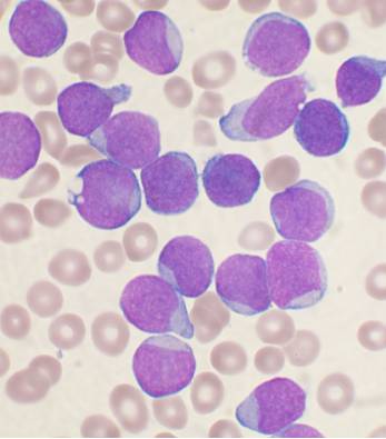
- Adults, median age ~65; risk factors: MDS, prior alkylating agents / topoisomerase-II inhibitors, radiation, Down syndrome
- Auer rods — crystallized azurophilic (primary) granules, strongly MPO+ — are effectively pathognomonic for AML; blasts also express CD13, CD33, CD117
- APL (M3): t(15;17) *PML-RARA* — hypergranular promyelocytes with numerous Auer rods ("faggot cells"); classically causes DIC
- APL is a medical emergency: start ATRA (all-trans retinoic acid) before genetic confirmation; definitive therapy adds arsenic trioxide. Watch for differentiation (ATRA) syndrome — fever, dyspnea, pulmonary edema/effusions — treat with dexamethasone
- Monocytic AML → gum (gingival) infiltration
- Favorable cytogenetics: t(8;21), inv(16)
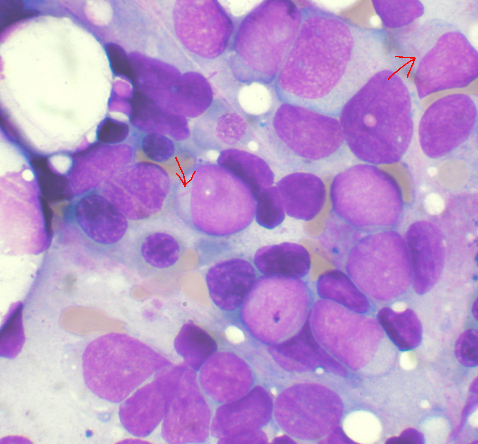
Vignette: A 38-year-old woman has 1 week of fatigue, gum bleeding, and spontaneous bruising. Labs: Hb 7.8, platelets 22k, WBC 2.5. Coags: ↑PT/PTT, ↓fibrinogen, ↑D-dimer. Smear: hypergranular promyelocytes packed with needle-like inclusions.
Diagnosis: Acute promyelocytic leukemia (APL / AML-M3) with DIC, driven by t(15;17).
Next best step: Start ATRA immediately — do not wait for cytogenetics — and support the coagulopathy (platelets, cryoprecipitate for fibrinogen, FFP as needed). ATRA drives promyelocyte differentiation, reversing the DIC. Definitive therapy adds arsenic trioxide.
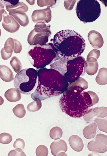
Vignette: A 4-year-old boy has weeks of fatigue, pallor, low-grade fevers, and limping / refusing to walk (bone pain). Exam: hepatosplenomegaly, diffuse lymphadenopathy, scattered petechiae. CBC: anemia, thrombocytopenia, WBC 40k with lymphoblasts; blasts are TdT+, CD10+.
Diagnosis: B-cell ALL — the most common childhood cancer.
Next best step: Bone marrow biopsy to confirm ≥20% blasts, with flow cytometry and cytogenetics. Treatment is multi-agent chemo plus CNS prophylaxis (intrathecal methotrexate) and tumor-lysis prophylaxis (hydration + rasburicase/allopurinol).
- Auer rods → AML (both start with A); an Auer rod = crystallized azurophilic granules (MPO+)
- APL = the "A" trio: APL / t(15;17) → ATRA + Arsenic
- TdT⁺ = immaTure lymphoblasts (ALL)
- Tumor lysis = "everything up but calcium": ↑K⁺, ↑phosphate, ↑uric acid, ↓Ca²⁺
- Philadelphia t(9;22) is bad in ALL — but add a TKI (imatinib)
Practice Heme/Onc now
Board-style questions, spaced-repetition flashcards, and a Socratic AI tutor — free to start.