Acute Kidney Injury: Prerenal, Intrinsic & Postrenal
A board-focused walkthrough of acute kidney injury that localizes each case to prerenal, intrinsic, or postrenal using BUN:Cr, FENa, and urine sediment, then drives the tested next-best-step and dialysis decisions. Emphasizes classic vignette buzzwords (muddy-brown casts, hyaline casts, eosinophiluria) and management reflexes.
Overview & KDIGO Definition
Acute kidney injury (AKI) is an abrupt (hours–days) fall in glomerular filtration causing retention of nitrogenous waste and dysregulation of fluid, electrolytes, and acid–base balance. By KDIGO criteria, diagnose AKI when serum creatinine rises ≥0.3 mg/dL within 48 h, ≥1.5× baseline within 7 days, or urine output is <0.5 mL/kg/h for ≥6 h.
Boards frame every case around three anatomic buckets:
- Prerenal — hypoperfusion with structurally intact tubules (most common overall; reversible if perfusion is restored promptly).
- Intrinsic — parenchymal injury to tubules, interstitium, glomeruli, or vessels; acute tubular necrosis (ATN) dominates.
- Postrenal — obstruction to urine outflow; must involve both kidneys (or a single functioning kidney) to raise creatinine.
The first move in any AKI vignette is to localize the lesion: use history, the BUN:Cr ratio, urinalysis with microscopy, and the FENa — then act.
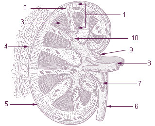
- Prerenal: ↓ effective circulating volume — hypovolemia (vomiting, diarrhea, hemorrhage, burns), heart failure, cirrhosis/hepatorenal syndrome, sepsis, bilateral renal artery stenosis.
- NSAIDs (afferent arteriole constriction) and ACE inhibitors/ARBs (efferent arteriole dilation) precipitate prerenal AKI; the combination is classically dangerous in renal artery stenosis.
- ATN = prolonged ischemia (from any prerenal state) or nephrotoxins: aminoglycosides, IV contrast, cisplatin, amphotericin B, and pigments (myoglobin in rhabdomyolysis, hemoglobin in hemolysis).
- AIN (acute interstitial nephritis) = allergic reaction to drugs — β-lactams, sulfonamides, NSAIDs, PPIs, rifampin, allopurinol; look for fever, rash, eosinophilia.
- Postrenal: BPH, bilateral stones, pelvic/cervical/prostate cancer, neurogenic bladder, retroperitoneal fibrosis.
- Emergencies needing dialysis: refractory hyperkalemia, acidosis, volume overload, uremic pericarditis/encephalopathy, dialyzable toxins.
Prerenal vs ATN vs Postrenal — The Urine Workup
| Parameter | Prerenal | Intrinsic (ATN) | Postrenal |
|---|---|---|---|
| BUN:Cr ratio | >20:1 | 10–15:1 | early >15, later ~10–15 |
| FENa | <1% | >2% | early <1%, late >2% |
| Urine Na (mEq/L) | <20 | >40 | varies |
| Urine osmolality | >500 (concentrated) | <350 (isosthenuric ~300) | <350 (late) |
| Sediment | bland / hyaline casts | muddy-brown granular casts, RTE cells | normal ± crystals |
| Mechanism | intact tubules avidly reabsorb Na/H₂O | tubular cell death + back-leak | ↑ tubular pressure |
Vignette: An 82-year-old man with 3 days of vomiting and diarrhea has dry mucous membranes, flat neck veins, and orthostatic hypotension. Labs: BUN 56, Cr 2.1 (baseline 1.0), BUN:Cr 27:1. Urine: Na 12 mEq/L, osmolality 640 mOsm/kg, FENa 0.4%, hyaline casts only.
Diagnosis: Prerenal azotemia from volume depletion — the tubules are intact and avidly conserving sodium and water.
Next best step: Isotonic IV fluids (normal saline or a balanced crystalloid) and hold nephrotoxins (NSAIDs, ACEi/ARB). Creatinine should improve within 24–72 h; failure to recover despite adequate volume signals progression to ischemic ATN — expect the FENa to climb >2% and muddy-brown casts to appear.
Intrinsic AKI — Match the Sediment to the Compartment
- ATN (tubules): muddy-brown granular and renal tubular epithelial cell casts. Three phases — initiation, maintenance (oliguric; watch hyperkalemia, metabolic acidosis, volume overload), and recovery (polyuric, risk of hypokalemia). Contrast-associated: Cr rises 24–48 h, peaks day 3–5, resolves in ~1 week.
- AIN (interstitium): WBC casts, sterile pyuria, urine eosinophils. The fever/rash/eosinophilia triad is often incomplete — suspect it whenever AKI follows a new drug. Stop the culprit ± corticosteroids.
- Glomerulonephritis: dysmorphic RBCs and RBC casts, proteinuria, hypertension, edema — the nephritic picture.
- Vascular: TTP/HUS (schistocytes, thrombocytopenia), scleroderma renal crisis (treat with an ACE inhibitor), and cholesterol (atheroembolic) disease after catheterization (livedo reticularis, blue toes, eosinophilia, low complement, appearing days–weeks later).
Vignette: A 74-year-old man with known BPH presents with 12 h of anuria, suprapubic fullness, and a palpable bladder. Cr is 3.4 (baseline 1.1).
Next best step: Place a bladder (Foley) catheter — it is both diagnostic and therapeutic. Drainage of a large residual volume confirms bladder-outlet obstruction and relieves it.
If catheter output is minimal, obtain renal ultrasound to look for bilateral hydronephrosis (upper-tract obstruction → needs nephrostomy or ureteral stent).
Anticipate post-obstructive diuresis after relief — monitor volume status and electrolytes closely. Key board point: unilateral obstruction with a normal contralateral kidney does not raise creatinine.
AEIOU — indications for emergent dialysis:
- Acidosis — severe, refractory metabolic acidosis
- Electrolytes — refractory hyperkalemia
- Intoxications — dialyzable toxins (I STUMBLED: Isopropanol, Salicylates, Theophylline, Uremia, Methanol, Barbiturates, Lithium, Ethylene glycol, Dabigatran)
- Overload — volume overload / refractory pulmonary edema
- Uremia — pericarditis, encephalopathy, or bleeding
Cast cheat sheet:
- RBC casts → glomerulonephRitis
- WBC casts → pyelonephritis / AIN
- muddy-brown granular casts → ATN
- fatty (Maltese-cross) casts → nephrotic syndrome
- broad, waxy casts → chronic kidney disease
- Prerenal: restore perfusion (IV fluids if hypovolemic; treat the HF/cirrhosis driver); stop NSAIDs/ACEi/ARB.
- ATN: supportive only — maintain euvolemia, correct electrolytes/acidosis, avoid nephrotoxins and repeat contrast. No drug reverses established ATN.
- AIN: discontinue the culprit drug; corticosteroids if renal function does not recover.
- Postrenal: relieve obstruction (Foley → ultrasound → nephrostomy/stent).
- Rhabdomyolysis: aggressive IV isotonic fluids; expect ↑CK, ↑K⁺, ↑PO₄, ↓Ca²⁺, and dipstick blood-positive with no RBCs on microscopy.
Hyperkalemia with ECG changes — order of action:
- IV calcium gluconate — membrane stabilization (does not lower K⁺)
- Insulin + glucose, albuterol, ± bicarbonate — shift K⁺ intracellularly
- Remove K⁺ — loop diuretic, GI cation binder, or dialysis
Practice Renal now
Board-style questions, spaced-repetition flashcards, and a Socratic AI tutor — free to start.