Acute Coronary Syndromes (STEMI, NSTEMI, Unstable Angina)
A board-focused walkthrough of acute coronary syndromes from plaque rupture to reperfusion, contrasting STEMI, NSTEMI, and unstable angina with the classic vignettes, ECG localization, next-best-step decisions, and the post-MI complication timeline. Emphasizes the high-yield twists boards test — RV infarct preload dependence, no lytics in NSTE-ACS, STEMI-equivalents (posterior MI, Sgarbossa-positive LBBB), and which therapies actually reduce mortality.
Pathophysiology: it's a supply problem
Acute coronary syndromes (ACS) are a spectrum of acute myocardial ischemia, almost always from rupture or erosion of an atherosclerotic plaque with a superimposed platelet-rich thrombus in an epicardial coronary artery. The degree and duration of occlusion define the syndrome:
- Unstable angina (UA) and NSTEMI: partial/subtotal occlusion → subendocardial ischemia. Distinguished only by troponin (positive = NSTEMI).
- STEMI: complete, persistent occlusion → transmural ischemia → myocyte necrosis.
Boards love the mechanism: a lipid-rich, thin-capped "vulnerable" plaque ruptures, exposing a thrombogenic core → tissue factor → thrombin → fibrin/platelet clot. Contrast with stable angina, a demand problem from a fixed plaque (pain on exertion, relieved by rest/nitrates). ACS is a supply problem from acute thrombosis — pain at rest, prolonged, crescendo. Cocaine causes ACS via coronary vasospasm (avoid beta-blockers → unopposed alpha-mediated vasoconstriction).
- Classic vignette: substernal chest pressure/tightness >20 min radiating to left arm or jaw, with diaphoresis, nausea, dyspnea; not relieved by rest or nitroglycerin.
- Levine sign: clenched fist held over the sternum.
- Atypical/silent presentations in diabetics, elderly, women, post-op patients — dyspnea, epigastric pain, syncope, or fatigue alone.
- Pain that is sharp, positional, or reproduced by palpation argues against ACS (but never fully excludes it).
- First test = 12-lead ECG within 10 minutes of arrival; repeat serial ECGs if symptoms persist or change.
- Troponin is the biomarker (high-sensitivity assays become detectable within ~1–3 h): peaks ~24 h, stays elevated 7–14 days. Draw serial troponins and look for a rise/fall pattern (a single elevated value can be chronic).
- CK-MB normalizes in 2–3 days → useful to detect re-infarction.
- UA = ischemic symptoms + negative troponin.

STEMI vs NSTEMI vs Unstable Angina
| Feature | Unstable Angina | NSTEMI | STEMI |
|---|---|---|---|
| Occlusion | Partial | Partial/subtotal | Complete |
| Ischemia | Subendocardial | Subendocardial | Transmural |
| ECG | ST depression / T-wave inversion or normal | ST depression / T-wave inversion | ST elevation in ≥2 contiguous leads (STEMI-equivalents: posterior MI, new LBBB by Sgarbossa) |
| Troponin | Negative | Positive | Positive |
| Reperfusion | No emergent reperfusion | No emergent reperfusion | Emergent PCI / lytics |
| Anti-thrombotic | ASA + P2Y12 + anticoagulant | ASA + P2Y12 + anticoagulant | ASA + P2Y12 + anticoagulant |
Stem: 58-year-old man, hypertension and smoking, with 45 min of crushing substernal chest pain radiating to the jaw, diaphoretic. ECG shows ST elevation in II, III, aVF with reciprocal ST depression in aVL (± I).
- Diagnosis: Inferior STEMI → culprit is usually the RCA.
- Next best step: aspirin (chewed) + activate the cath lab for primary PCI.
Board twist: obtain right-sided leads (V4R). If RV infarct (hypotension, clear lungs, elevated JVD, ST elevation in V4R) the patient is preload-dependent → give IV fluids and avoid nitrates and morphine (they drop preload → profound hypotension). Also watch for bradycardia/AV block — the RCA supplies the SA node (~60%) and AV node (~90%).
Stem: 67-year-old diabetic woman with 30 min of rest chest pressure, now resolved. ECG shows 1 mm ST depressions in V4–V6. Serial troponin returns elevated.
- Diagnosis: NSTEMI.
- Management: dual antiplatelet (aspirin + a P2Y12 inhibitor) + anticoagulation (heparin) + anti-ischemic therapy (beta-blocker, nitrates) + high-intensity statin. NO fibrinolytics — they are harmful in NSTE-ACS.
- Risk-stratify with TIMI or GRACE scores.
- High-risk features (refractory/ongoing pain, dynamic ECG changes, rising troponin, hemodynamic or electrical instability, GRACE >140) → early invasive angiography within 24 h. Very-high-risk/unstable → immediate cath.
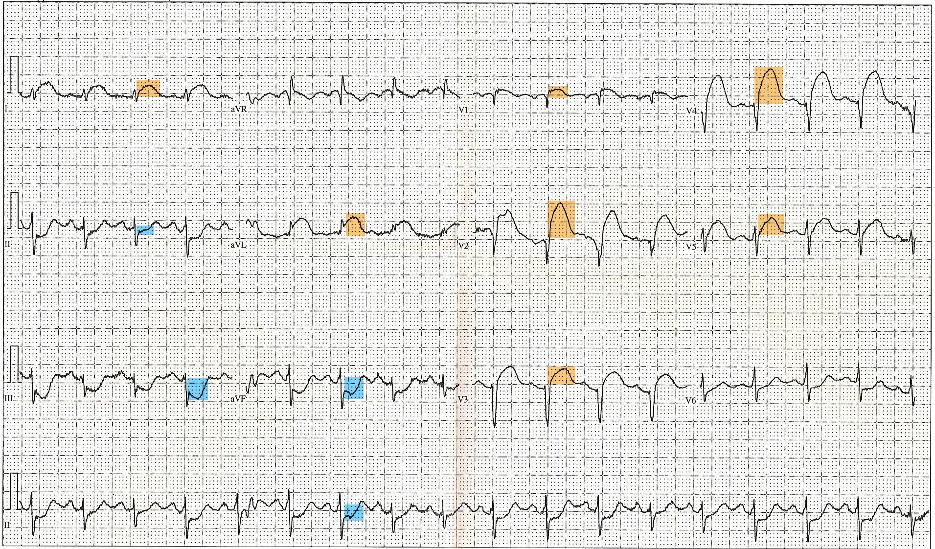
- STEMI is a reperfusion emergency — restore flow fast.
- Primary PCI preferred: first-medical-contact–to-device ≤90 min (≤120 min if transfer needed).
- Fibrinolysis (e.g., tenecteplase) if timely PCI is unavailable (anticipated FMC-to-device >120 min): door-to-needle ≤30 min, best within 12 h of symptom onset and no contraindications (active bleeding, any prior intracranial hemorrhage, ischemic stroke within 3 months, severe uncontrolled HTN).
- STEMI-equivalents — also mandate emergent reperfusion: posterior MI (ST depression V1–V3 + tall R waves → confirm with posterior leads V7–V9), new LBBB meeting Sgarbossa criteria, and de Winter T-waves (upsloping ST depression + tall symmetric T's = proximal LAD occlusion).
- Immediate meds: aspirin 162–325 mg chewed (mortality benefit) + P2Y12 inhibitor (ticagrelor/prasugrel/clopidogrel) + anticoagulation (heparin).
- Adjuncts: high-intensity statin; oral beta-blocker within 24 h (hold if hypotension, bradycardia, or signs of acute HF/cardiogenic shock — early IV beta-blockade caused shock in COMMIT); ACE inhibitor (esp. anterior MI, low EF, HF).
- Oxygen only if SpO₂ <90% — routine O₂ has no benefit.
MONA-B — the classic initial ACS mnemonic, but boards test the caveats more than the letters:
- Morphine — for refractory pain only; it can mask ongoing ischemia and delays P2Y12 absorption (use sparingly).
- Oxygen — only if SpO₂ <90%.
- Nitrates — contraindicated in RV infarct, hypotension, or recent PDE5 inhibitor use (sildenafil <24 h, tadalafil <48 h).
- Aspirin — the mortality-reducer; give to everyone without a contraindication.
- Beta-blocker — hold in acute HF/cardiogenic shock.
Key point: of MONA, only Aspirin — plus reperfusion, high-intensity statins, ACE inhibitors, and beta-blockers — actually improves survival. Morphine, oxygen, and nitrates are largely symptomatic.
Post-MI complications timeline (STEP 1 favorite)
| Timing | Complication | Key clue |
|---|---|---|
| Minutes–hours | Ventricular fibrillation | #1 cause of pre-hospital death |
| Hours | Cardiogenic shock | Large anterior MI, hypotension, pulmonary edema |
| 1–3 days | Fibrinous pericarditis | Friction rub, pleuritic pain (post-transmural) |
| 3–5 days | Papillary muscle rupture | New MR murmur, flash pulmonary edema (posteromedial = single PDA supply) |
| 3–5 days | Septal (VSD) rupture | New holosystolic murmur, O₂ step-up RA→RV |
| 5–14 days | Free wall rupture | Hemopericardium → tamponade, PEA, sudden death |
| Weeks–months | LV aneurysm / Dressler | Persistent ST elevation; late autoimmune pericarditis + effusion |
Practice Cardiology now
Board-style questions, spaced-repetition flashcards, and a Socratic AI tutor — free to start.