The Acute Abdomen: Differential & Workup
A boards-focused approach to the acute abdomen: use pain type, peritoneal signs, and hemodynamics to separate surgical emergencies from medical mimics, then apply the classic buzzword → next-best-step reflexes (unstable + peritonitis = OR; βhCG first; free air = perforation; pain out of proportion = mesenteric ischemia).
Framing the Acute Abdomen
An acute abdomen is the sudden onset of severe abdominal pain (usually < 24–48 h) that may signal a surgical emergency. Your first job is not to name the disease but to answer one question: does this patient need the operating room now?
Pain type localizes the problem. Visceral pain is dull, poorly localized, and midline (stretch of a hollow viscus). Parietal (somatic) pain is sharp, well-localized, and worse with movement or coughing (peritoneal irritation). The migration of periumbilical → RLQ pain in appendicitis is the classic visceral-to-parietal shift.
Peritoneal signs — involuntary guarding, rigidity ("board-like" abdomen), and rebound tenderness — mean peritonitis until proven otherwise. The single most important board reflex: a hemodynamically unstable patient with peritonitis goes to the OR — do NOT delay for CT.
- Unstable + peritonitis = laparotomy now. Imaging is for the stable patient.
- βhCG first in every reproductive-age woman with abdominal pain — it can flip the entire differential (ectopic) and must precede CT/radiation.
- Free air under the diaphragm on upright CXR = perforated viscus (classically perforated peptic ulcer) → surgery.
- Pain out of proportion to exam = acute mesenteric ischemia until disproven.
- Charcot triad (RUQ pain, fever, jaundice) = ascending cholangitis → urgent biliary decompression.
- CT abdomen/pelvis with IV contrast is the workhorse in stable patients; RUQ ultrasound is first-line for biliary disease and in children/pregnant patients.
- Get an ECG — inferior MI and DKA both masquerade as epigastric pain.
- The elderly, diabetic, and immunosuppressed have blunted exams and higher mortality; keep a low threshold for imaging.
Rigid, board-like abdomen + free air under the diaphragm (sudden severe epigastric pain; NSAID use or H. pylori). → Perforated peptic ulcer. NEXT BEST STEP: resuscitate (IVF, IV PPI, broad-spectrum antibiotics, NG tube) and go to the OR for repair (Graham omental patch). Upright CXR confirms free air; CT if equivocal.
Elderly smoker, hypotension, sudden severe back/flank pain, pulsatile abdominal mass. → Ruptured AAA. NEXT BEST STEP: if unstable → immediate OR; if stable and diagnosis uncertain → CT angiography. Never wait on imaging for a crashing patient.
Reproductive-age woman, amenorrhea, syncope, adnexal tenderness, +βhCG. → Ruptured ectopic pregnancy. NEXT BEST STEP: βhCG + transvaginal ultrasound; if unstable, straight to surgery (salpingectomy) — do not delay for further imaging.
Pain Location → Differential → First Test
| Location | Think first | Initial best test |
|---|---|---|
| RUQ | Cholecystitis, cholangitis, hepatitis | RUQ ultrasound |
| Epigastric | Pancreatitis, PUD/perforation, inferior MI | Lipase, upright CXR, ECG |
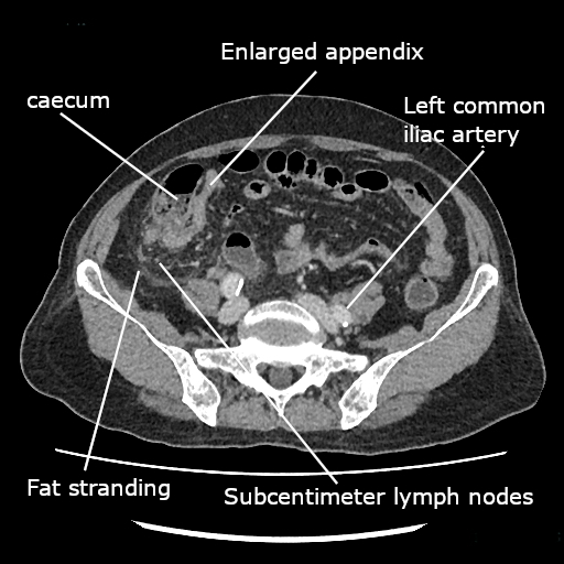
| RLQ | Appendicitis, ectopic, ovarian torsion, Crohn's | CT (US if child/pregnant) + βhCG |
| LLQ | Diverticulitis, ectopic, ovarian torsion | CT abd/pelvis + βhCG |
| Flank/back | Nephrolithiasis, pyelonephritis, ruptured AAA | Non-con CT (stone); US/CTA (AAA) |
| Diffuse | Perforation, mesenteric ischemia, SBO, DKA | Upright CXR, lactate, CT ± CTA |

Charcot triad → ascending cholangitis: RUQ pain + fever + jaundice. Add hypotension + altered mental status = Reynolds pentad (suppurative cholangitis — emergency decompression).
MANTRELS (Alvarado score for appendicitis):
- Migration of pain to RLQ
- Anorexia
- Nausea/vomiting
- Tenderness in RLQ
- Rebound tenderness
- Elevated temperature
- Leukocytosis
- Shift of WBCs left (neutrophilia)
Appendicitis special signs: Rovsing (LLQ palpation → RLQ pain), Psoas (RLQ pain on hip extension), Obturator (pain on internal rotation of the flexed hip).
Pain out of proportion to a benign exam + atrial fibrillation + lactic acidosis (± later bloody diarrhea). → Acute mesenteric ischemia (SMA embolus). NEXT BEST STEP: CT angiography; resuscitate, anticoagulate, and surgical embolectomy/revascularization with resection of necrotic bowel.
RUQ pain + fever + jaundice (Charcot triad) after gallstone disease. → Ascending cholangitis. NEXT BEST STEP: IV fluids + antibiotics, then urgent biliary decompression by ERCP (RUQ US + cholestatic LFTs support it).
Elderly, constipated/institutionalized patient; "coffee-bean" (inverted-U) loop on x-ray. → Sigmoid volvulus. NEXT BEST STEP: if no peritonitis, endoscopic detorsion (flexible sigmoidoscopy) then elective sigmoidectomy; peritonitis/ischemia → surgery.
Prior abdominal surgery, colicky pain, distension, vomiting, obstipation; air-fluid levels/dilated loops on x-ray. → Small bowel obstruction (adhesions). NEXT BEST STEP: NPO, NG decompression, IV fluids; CT to find the transition point/strangulation. Fever, tachycardia, leukocytosis, or peritonitis = strangulation → surgery.
- Resuscitate first: two large-bore IVs, crystalloid, type & screen, correct coagulopathy — ABCs before diagnostics.
- Labs: CBC, BMP, LFTs, lipase, lactate, UA, βhCG, coags. Lactate flags ischemia/necrosis.
- Upright CXR (or abdominal series): free air under the diaphragm → perforation.
- CT abdomen/pelvis with IV contrast: workhorse for the stable patient (appendicitis, diverticulitis, SBO; add CTA for ischemia).
- Ultrasound first for suspected biliary disease and in children, pregnant patients, and gynecologic pathology (ovarian torsion → pelvic US with Doppler); bedside US measures the aorta for AAA and detects free intraperitoneal fluid (FAST in trauma).
- HIDA scan if RUQ ultrasound is equivocal for cholecystitis.
- Do NOT perform colonoscopy in acute diverticulitis (perforation risk); scope electively ~6 weeks later to exclude malignancy.
Surgical Emergencies at a Glance
| Entity | Classic buzzword | Best diagnostic test | Definitive management |
|---|---|---|---|
| Perforated peptic ulcer | Rigid abdomen, free air under diaphragm | Upright CXR; CT if equivocal | Laparotomy + Graham patch |
| Ruptured AAA | Hypotension, pulsatile mass, back pain | Unstable → OR; stable → CTA | Open/endovascular repair |
| Ruptured ectopic | Amenorrhea, +βhCG, shock | βhCG + transvaginal US | Surgery (salpingectomy) |
| Acute mesenteric ischemia | Pain out of proportion, A-fib, ↑lactate | CT angiography | Embolectomy + resect dead bowel |
| Ascending cholangitis | Charcot triad / Reynolds pentad | RUQ US + cholestatic LFTs | IVF + abx → ERCP decompression |
| Strangulated SBO | Prior surgery/hernia, air-fluid levels | CT (transition point, closed loop) | Resuscitate + surgery |
| Sigmoid volvulus | "Coffee-bean" sign, elderly | Abd x-ray / CT | Endoscopic detorsion → sigmoidectomy |
Pitfalls & Special Populations
Atypical presentations kill. The elderly often lack fever, leukocytosis, and a rigid abdomen even with perforation or ischemia — yet carry far higher mortality; image early and trust vitals over the exam. Diabetics and the immunosuppressed (steroids, chemotherapy) mount blunted inflammatory responses and can hide an advanced process behind a soft belly.
Always check a glucose and an ECG: DKA and inferior MI both present with epigastric pain and can mimic — or accompany — a surgical abdomen. In any reproductive-age woman, ectopic pregnancy stays on the differential until βhCG and pelvic ultrasound say otherwise. When the exam is equivocal but the patient looks sick, serial exams and early surgical consultation beat premature reassurance.
Practice Surgery now
Board-style questions, spaced-repetition flashcards, and a Socratic AI tutor — free to start.