Abdominal Wall Hernias
A board-focused walkthrough of abdominal wall hernias: the anatomy that separates indirect, direct, and femoral groin hernias, the exam landmarks that localize them, and the reducible → incarcerated → strangulated triage that drives every next-best-step decision.
The Big Picture
An abdominal wall hernia is protrusion of a viscus (usually bowel or omentum) through a defect in the wall that normally contains it. The clinical stakes fall along a three-state continuum. A reducible hernia can be pushed back into the abdomen. An incarcerated hernia is stuck (irreducible) and may obstruct. A strangulated hernia has a compromised blood supply — an ischemic surgical emergency signaled by exquisite tenderness, erythema over the sac, and systemic toxicity (fever, tachycardia, leukocytosis).
Boards test three things: the anatomy that distinguishes hernia types, the landmarks that localize them on exam, and — most importantly — the NEXT BEST STEP once complications appear. Inguinal hernias alone account for ~75% of all abdominal wall hernias, and the indirect inguinal hernia is the single most common type in both sexes and at all ages.
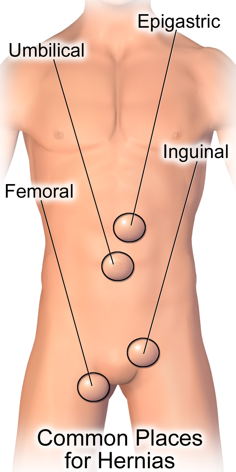
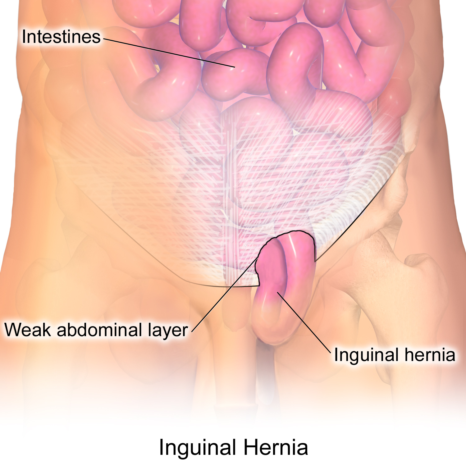
- Indirect inguinal = most common hernia overall; passes through the deep ring LATERAL to the inferior epigastric vessels, follows the spermatic cord, and can reach the scrotum; cause is a patent processus vaginalis (congenital)
- Direct inguinal = bulges through Hesselbach triangle, MEDIAL to the inferior epigastric vessels; acquired weakness of the transversalis fascia in older men; rarely enters the scrotum
- Hesselbach triangle borders: inferior epigastric vessels (superolateral), lateral border of rectus abdominis (medial), inguinal ligament (inferior)
- Femoral = through the femoral canal, BELOW the inguinal ligament, MEDIAL to the femoral vein; more common in women; highest risk of incarceration/strangulation
- Pubic tubercle rule: inguinal hernias sit superomedial; femoral hernias sit inferolateral to it
- Groin hernias are clinical diagnoses — image (ultrasound first) only when the exam is equivocal
Indirect vs Direct vs Femoral
| Feature | Indirect inguinal | Direct inguinal | Femoral |
|---|---|---|---|
| Vs. inf. epigastric vessels | Lateral | Medial | Below inguinal lig. |
| Route | Deep ring → cord → scrotum | Hesselbach triangle | Femoral canal |
| Typical patient | Any age; #1 overall; infants/young | Older men | Women |
| Cause | Patent processus vaginalis (congenital) | Acquired fascial weakness | Acquired |
| Enters scrotum | Often | Rarely | No |
| Strangulation risk | Moderate | Low | High |
MDs don't LIE — relative to the inferior epigastric vessels:
- Medial = Direct
- Lateral = Indirect
Pubic tubercle localizer — rest a finger on the pubic tubercle:
- Inguinal hernia bulges above and medial (superomedial)
- Femoral hernia bulges below and lateral (inferolateral)
Internal-ring occlusion test — reduce the hernia, occlude the deep internal ring (just above the midpoint of the inguinal ligament), and have the patient cough: if the bulge is controlled, it is indirect; if it still protrudes medially, it is direct. (Classic exam concept — clinically imperfect and not used to decide management.)
Vignette: A 68-year-old man has 8 hours of a painful, firm groin bulge he can no longer push back in, with nausea, vomiting, and obstipation. Temp 38.4°C, HR 112. The overlying skin is erythematous and the mass is exquisitely tender; labs show leukocytosis.
Diagnosis: Strangulated inguinal hernia — incarceration + ischemia + systemic toxicity.
Next best step: Emergent surgical exploration with resection of any nonviable bowel. Do NOT attempt manual reduction — pushing potentially necrotic bowel back into the abdomen risks perforation and peritonitis. IV fluids, NG decompression, and antibiotics are adjuncts, not substitutes for the OR.
Contrast: a reducible or freshly incarcerated hernia without strangulation signs (soft, non-tender, no toxicity) may get a gentle reduction attempt followed by repair.
Vignette: A 72-year-old thin woman has crampy abdominal pain, vomiting, and distension. Exam shows a small, tender bulge below and lateral to the right pubic tubercle, inferior to the inguinal ligament. Abdominal films show dilated small-bowel loops with air-fluid levels.
Diagnosis: Incarcerated femoral hernia causing small-bowel obstruction. Femoral hernias are more common in women and carry the highest strangulation risk of the groin hernias because of the narrow, rigid femoral ring.
Next best step / principle: Surgical repair — here urgent given the obstruction. Because of their high complication rate, femoral hernias are repaired even when asymptomatic. Watchful waiting is acceptable for minimally symptomatic inguinal hernias, but never for femoral hernias.
- Umbilical: infants — usually closes spontaneously by age 4–5 (repair if persistent beyond that, large, or incarcerated); adults — from raised intra-abdominal pressure (obesity, ascites, pregnancy)
- Incisional (ventral): at a prior surgical scar; risk factors obesity, wound infection
- Spigelian: through the linea semilunaris (Spigelian fascia); lateral and often occult — high incarceration risk
- Obturator: elderly thin women; Howship–Romberg sign = medial thigh/knee pain from obturator nerve compression
- Richter: only the antimesenteric bowel wall herniates — can strangulate without obstruction
- Littre: contains a Meckel diverticulum
- Amyand: contains the appendix
- Sliding: a retroperitoneal organ (colon, bladder) forms part of the sac wall
Management Logic the Boards Reward
Triage first by complication status. Strangulation (severe pain, tenderness, skin change, systemic toxicity) → straight to the OR, no reduction. Incarceration without strangulation → gentle reduction attempt, then repair. Reducible hernias → elective repair, usually tension-free mesh (open or laparoscopic); watchful waiting is reasonable only for minimally symptomatic inguinal hernias in adults.
Two absolutes: femoral hernias are always repaired, and every strangulated hernia is an emergency. In children, umbilical hernias are observed to age 4–5, while an indirect inguinal hernia from a persistently patent processus vaginalis does not resolve and is repaired to prevent incarceration. When a groin mass is diagnostically unclear, ultrasound is the first imaging test; reserve CT for obstruction or occult/complex cases.
Practice Surgery now
Board-style questions, spaced-repetition flashcards, and a Socratic AI tutor — free to start.