Normal·Special Senses·Eye
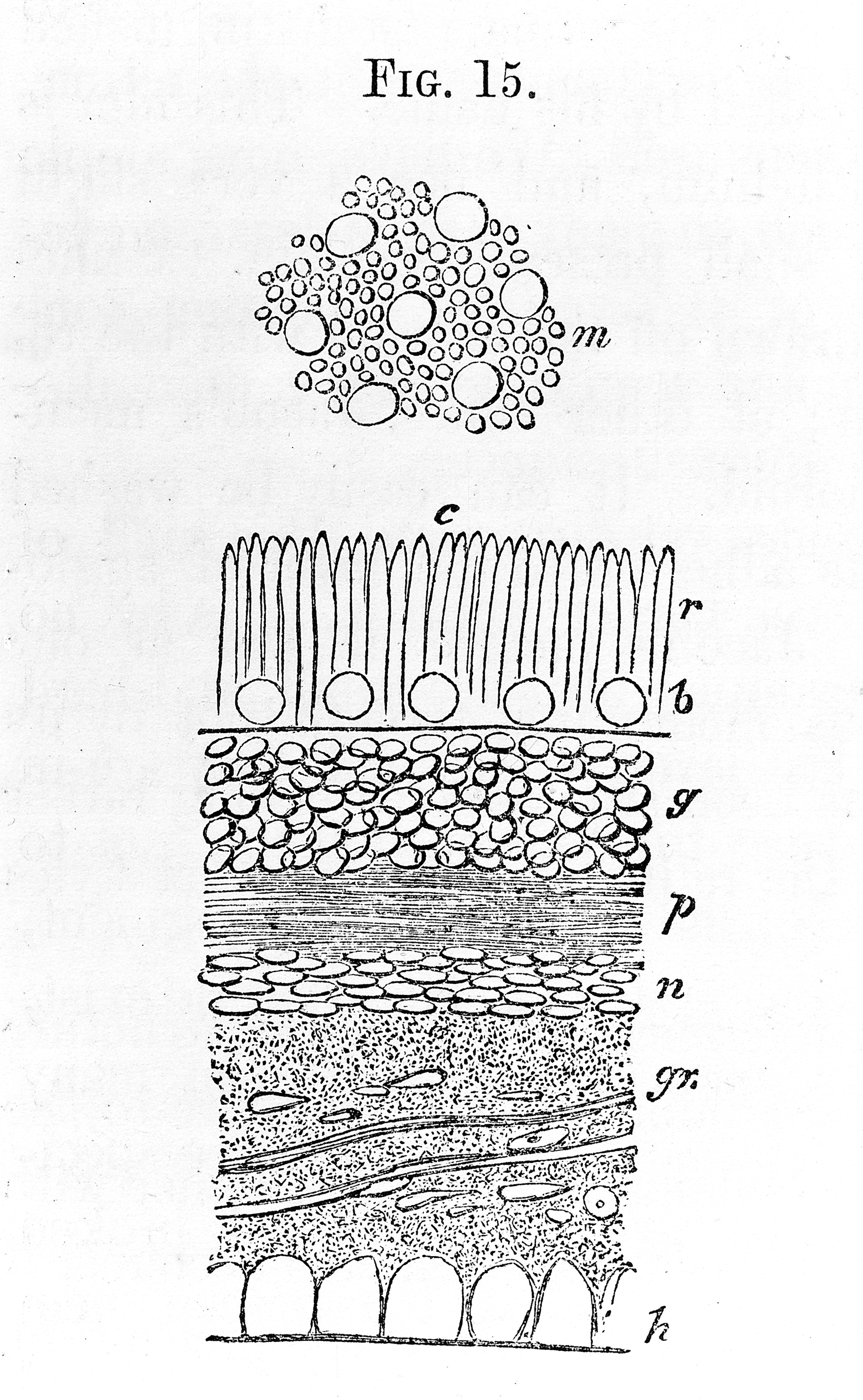
Retina — vertical section showing all layers
Stain: H&E·Magnification: 40x·Tissue: Neural retina + RPE·5 labeled regions
Wikimedia Commons · File:Vertical_section_of_the_human_retina._Wellcome_M0011269.jpg · https://commons.wikimedia.org/wiki/File:Vertical_section_of_the_human_retina._Wellcome_M0011269.jpg (CC-licensed)
Open the interactive viewer
Sign up free to pan/zoom up to 10×, tap each labeled marker for the structure's description and clinical context, and switch to quiz mode to test yourself on every region.
Create free accountDescription
Light passes inside → out: ganglion cells → bipolar cells → photoreceptors → RPE. The layered nuclei alternate with synaptic (plexiform) layers. Detachment occurs at the embryonic cleft between neural retina + RPE.
Labeled regions (5)
- 1Ganglion cell layer
Cell bodies of output neurons whose axons form the optic nerve. Damaged in glaucoma (cup-disc ratio↑).
- 2Inner nuclear layer
Bipolar, horizontal, amacrine, Müller cell bodies. Intermediate processing.
- 3Outer nuclear layer
Photoreceptor cell bodies (rods + cones). Far thicker than inner — rods outnumber cones ~20:1 outside the fovea.
- 4Photoreceptor outer segments
Stacks of disc membranes containing rhodopsin / opsins. Phagocytosed daily by RPE.
- 5Retinal pigment epithelium (RPE)
Single pigmented layer outermost; recycles 11-cis retinal + phagocytoses disc tips. Drusen accumulate here in dry AMD.